

Endogenous leukemia inhibitory factor protects photoreceptor cells against light-induced degeneration
- PMID: 19693290
- PMCID: PMC2728564
Endogenous leukemia inhibitory factor protects photoreceptor cells against light-induced degeneration
Abstract
Purpose: Expression of leukemia inhibitory factor (LIF) by a subset of Müller glia cells has recently been implicated in an endogenous survival response to photoreceptor injury in a model of inherited retinal degeneration. To investigate whether such a LIF-controlled survival pathway might be commonly induced upon photoreceptor injury independently of the nature of the toxic stimulus, we analyzed the role of LIF during light-induced retinal degeneration.
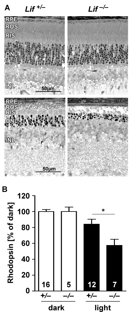
Methods: Lif(+/-) and Lif(-/-) mice were exposed to 15,000 lx of white light for 2 h. Retinal morphology and rhodopsin content were analyzed nine days after light exposure. Gene expression studies were done using real-time PCR. Protein levels were determined by western blotting using specific antibodies.
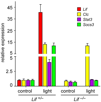
Results: A lack of LIF reduced survival of photoreceptor cells after light exposure. In the absence of LIF several genes encoding molecules involved in the Janus kinase/signal transducer and activator of transcription (Jak/STAT) signaling pathway were not activated after light exposure. Presence or absence of LIF did not affect AKT (also known as protein kinase B, PKB) signaling and had only a mild effect on extracellular regulated kinase (ERK) phosphorylation. Stress-induced glial fibrillary acidic protein (GFAP) induction was minimal in the absence of LIF.
Conclusions: Our results suggest that increased retinal expression of LIF is a general response to photoreceptor injury. Independent of the nature of the toxic insult (gene mutation, light), LIF may activate an endogenous rescue pathway that protects viable photoreceptor cells, leading to an increased photoreceptor survival in the stressed retina. This defense system may depend on the Jak/STAT pathway and may involve endothelin 2 (EDN2) but not (or only minimally) AKT and ERK1,2 signaling.
Figures


References
-
- Cook HL, Patel PJ, Tufail A. Age-related macular degeneration: diagnosis and management. Br Med Bull. 2008;85:127–49. - PubMed
-
- Kamphuis W, Dijk F, Bergen AA. Ischemic preconditioning alters the pattern of gene expression changes in response to full retinal ischemia. Mol Vis. 2007;13:1892–901. - PubMed
-
- Roth S. Endogenous neuroprotection in the retina. Brain Res Bull. 2004;62:461–6. - PubMed
-
- Grimm C, Wenzel A, Groszer M, Mayser H, Seeliger M, Samardzija M, Bauer C, Gassmann M, Reme CE. HIF-1-induced erythropoietin in the hypoxic retina protects against light-induced retinal degeneration. Nat Med. 2002;8:718–24. - PubMed
Publication types
MeSH terms
Substances
LinkOut - more resources
Full Text Sources
Molecular Biology Databases
Miscellaneous