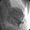
Successful aneurysmectomy of a congenital apical left ventricular aneurysm
- PMID: 19693309
- PMCID: PMC2720295
Successful aneurysmectomy of a congenital apical left ventricular aneurysm
Abstract
Congenital apical left ventricular aneurysm is a rare clinical entity that is different from congenital left ventricular diverticulum. This aneurysm usually occurs as an isolated anomaly. Its clinical presentation varies, and it is usually diagnosed by exclusion. Herein, we report the case of a 54-year-old man who experienced progressively increasing symptoms of congestive cardiac failure. Through the use of contrast echocardiography and angiocardiography, and upon histopathologic examination, he was diagnosed to have a congenital apical left ventricular aneurysm. He was successfully treated by means of left ventricular aneurysmectomy. We discuss the process of diagnosis and surgical correction of the aneurysm, and we briefly review the pertinent medical literature.
Keywords: Coronary aneurysm/surgery; diagnosis, differential; echocardiography; heart aneurysm/congenital/diagnosis/radiography/surgery; heart ventricles/abnormalities/radiography.
Figures


References
-
- Papagiannis J, Van Praagh R, Schwint O, D'Orsogna L, Qureshi F, Reynolds J, et al. Congenital left ventricular aneurysm: clinical, imaging, pathologic, and surgical findings in seven new cases. Am Heart J 2001;141(3):491–9. - PubMed
-
- Krasemann T, Gehrmann J, Fenge H, Debus V, Loeser H, Vogt J. Ventricular aneurysm or diverticulum? Clinical differential diagnosis. Pediatr Cardiol 2001;22(5):409–11. - PubMed
-
- Swyer AJ, Mauss IH, Rosenblatt P. Congenital diverticulosis of left ventricle. Am J Dis Child 1950;79(1):111–4. - PubMed
Publication types
MeSH terms
Substances
LinkOut - more resources
Full Text Sources
Medical
