Review
. 2010 Jan;339(1):247-57.
doi: 10.1007/s00441-009-0844-4. Epub 2009 Aug 20.

Collagens

Affiliations
Review

Collagens

Marion K Gordon et al. Cell Tissue Res. 2010 Jan.

Abstract

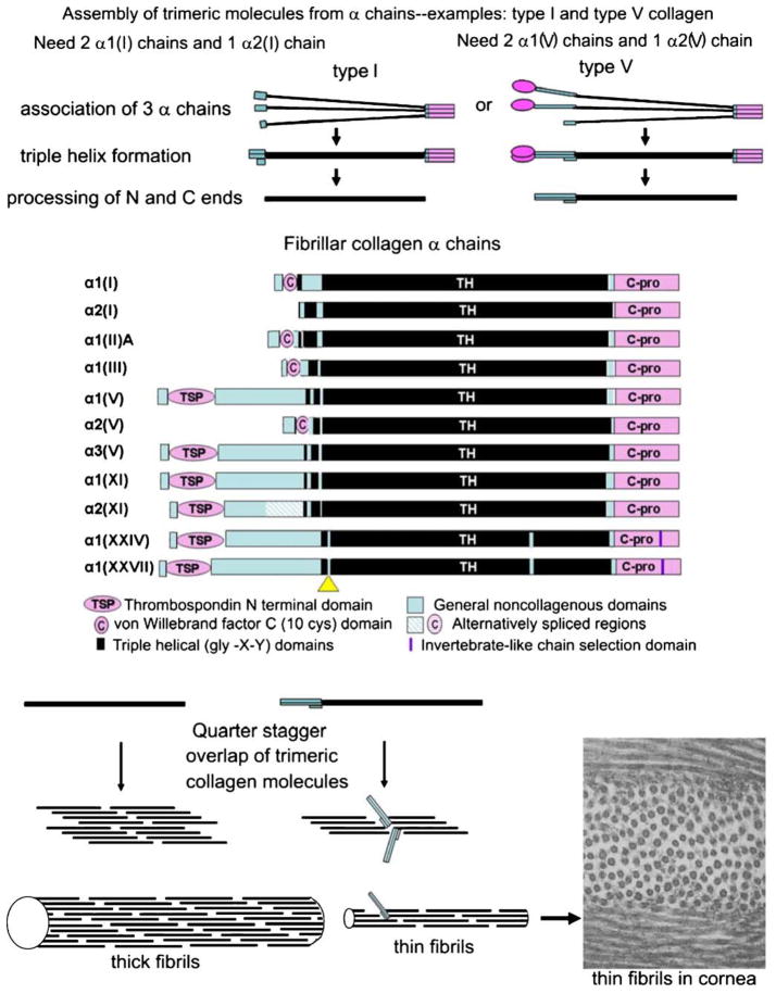
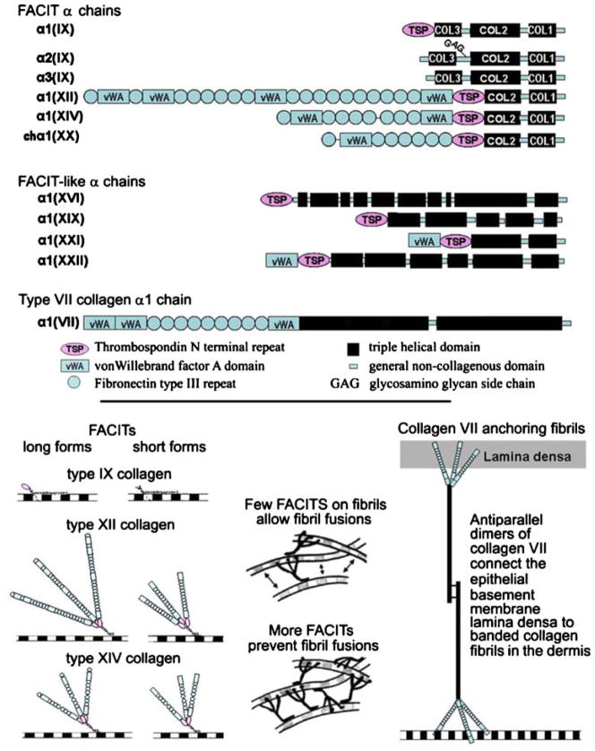
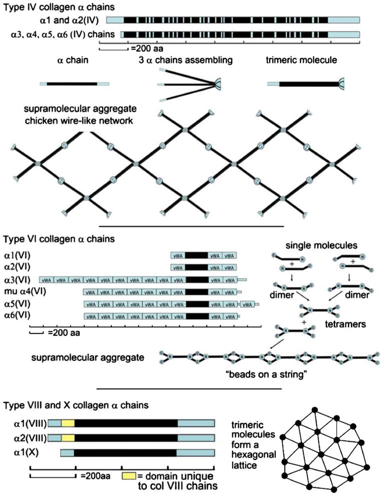
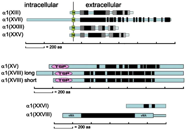
The collagens represent a family of trimeric extracellular matrix molecules used by cells for structural integrity and other functions. The three alpha chains that form the triple helical part of the molecule are composed of repeating peptide triplets of glycine-X-Y. X and Y can be any amino acid but are often proline and hydroxyproline, respectively. Flanking the triple helical regions (i.e., Col domains) are non-glycine-X-Y regions, termed non-collagenous domains. These frequently contain recognizable peptide modules found in other matrix molecules. Proper tissue function depends on correctly assembled molecular aggregates being incorporated into the matrix. This review highlights some of the structural characteristics of collagen types I-XXVIII.


Figures

Fig. 1
Fig. 1
Assembly of α chains into trimeric collagen molecules and then of molecules into fibrils. Top The folding of three chains into a single molecule. Middle Fibrillar collagen α chains showing domain structures. Scale is approximate (C-pro C-propeptide domain, TH gly-X-Y repeating region). The yellow triangle is a short non-gly-X-Y domain in collagens XXIV and XXVII and has been interpreted in two ways: as a separation between the minor and major helices in collagen XXIV, and as an interruption in the major (and only) triple helix in collagen XXVII. Bottom Perfect quarter stagger overlaps result when no bulky groups protrude from the fibril surface. Bottom right Electron micrograph of collagen fibrils in the rabbit cornea. The thin diameters (~25 nm) are the result of fibrils being heterotypic structures composed of ~80% type I and 20% type V collagen. ×60,000
Fig. 2
Fig. 2
Collagens associated with banded fibrils. Top FACIT α chains, FACIT-like α chains, and the type VII α1 chain. Domains are indicated below. Scale is approximate. Bottom left Long and short forms of types IX, XII, and XIV collagen on the surface of a fibril. Bottom middle The presence of a few FACITs allows the collagen fibrils to come into contact (double-headed arrows) and to fuse forming larger diameter structures. More FACITs associated with the fibril surface hinders fibril fusions. This has been shown recently for type XIV collagen. Bottom right A collagen VII antiparallel dimer functioning as a “rivet” linking the epithelial basement membrane with the banded fibrils of the dermis
Fig. 3
Fig. 3
Network-forming collagens. Top Representative type IV collagen chains. The chicken wire supramolecular aggregate of type IV collagen is shown below. Middle Type VI collagen chains, including the murine α4, which does not have a human counterpart. Single molecules forming dimers, and dimers interacting to become tetramers are presented right. The tetramers aggregate end to end. Bottom The α chains for short chain collagens, types VIII and X (yellow boxes domains unique to the collagen VIII α chains, blue boxes conserved non-collagenous domains, black boxes collagenous domains that are also conserved between collagen VIII and X). A drawing of a hexagonal lattice, a supramolecular aggregate that these collagens can assume, is shown right
Fig. 4
Fig. 4
Transmembrane, endostatin precursor, and other collagens. The α chains for each collagen are shown (black bars collagenous domains, blue bars NC domains, TM in green boxes transmembrane domains). In the transmembrane group, matching geometric patterns in collagens XIII, XXIII, and XXV indicate conserved regions (TSP thrombospondin N-terminal module, vWA von Willebrand factor A domains)

References

    1. Anderson S, SundarRaj S, Fite D, Wessel H, SundarRaj N. Developmentally regulated appearance of spliced variants of type XII collagen in the cornea. Invest Ophthalmol Vis Sci. 2000;41:55–63. - PubMed
    1. Ansorge HL, Meng X, Zhang G, Veit G, Sun M, Klement JF, Beason DP, Soslowsky LJ, Koch M, Birk DE. Type XIV collagen regulates fibrillogenesis: premature collagen fibril growth and tissue dysfunction in null mice. J Biol Chem. 2009;284:8427–8438. - PMC - PubMed
    1. Aumailley M, Has C, Tunggal L, Bruckner-Tuderman L. Molecular basis of inherited skin-blistering disorders, and therapeutic implications. Expert Rev Mol Med. 2006;8:1–21. - PubMed
    1. Bader HL, Keene DR, Charvet B, Veit G, Driever W, Koch M, Ruggiero F. Zebrafish collagen XII is present in embryonic connective tissue sheaths (fascia) and basement membranes. Matrix Biol. 2009;28:32–43. - PubMed
    1. Banyard J, Bao L, Zetter BR. Type XXIII collagen, a new transmembrane collagen identified in metastatic tumor cells. J Biol Chem. 2003;278:20989–20994. - PubMed

Publication types

LinkOut - more resources