Review
. 2010 Mar;19 Suppl 1(Suppl 1):S52-65.
doi: 10.1007/s00586-009-1124-4. Epub 2009 Aug 20.

Endoscopic surgery on the thoracolumbar junction of the spine

Affiliations
Review

Endoscopic surgery on the thoracolumbar junction of the spine

Rudolf Beisse. Eur Spine J. 2010 Mar.

Abstract

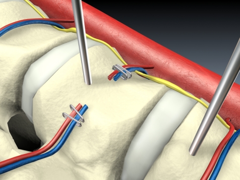
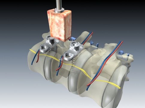
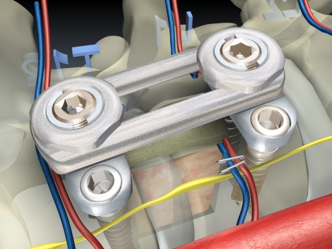
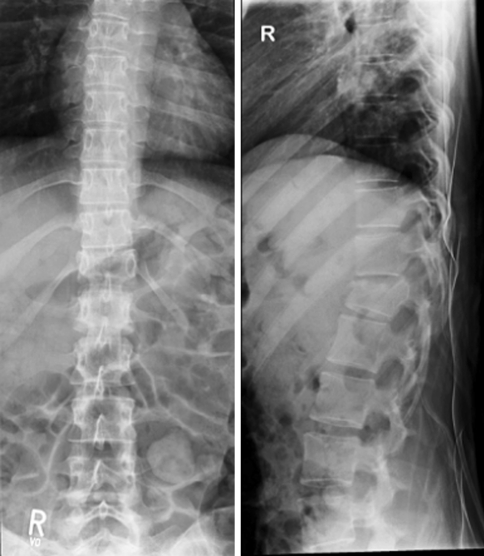
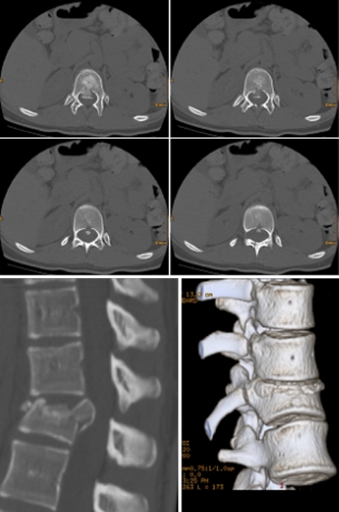
The thoracolumbar junction is the section of the truncal spine most often affected by injuries. Acute instability with structural damage to the anterior load bearing spinal column and post-traumatic deformity represent the most frequent indications for surgery. In the past few years, endoscopic techniques for these indications have partially superseded the open procedures, which are associated with high access morbidity. The particular position of this section of the spine, which lies in the border area between the thoracic and abdominal cavities, makes it necessary in most cases to partially detach the diaphragm endoscopically in order to expose the operation site, and this also provides access to the retroperitoneal section of the thoracolumbar junction. A now standardised operating technique and instruments and implants specially developed for the endoscopic procedure, from angle stable plate and screw implants to endoscopically implantable vertebral body replacements, have gradually opened up the entire spectrum of anterior spine surgery to endoscopic techniques.


Figures

Fig. 1
Fig. 1
Standard set up for endoscopic spine surgery. The patient is placed in a true lateral position; four portals are positioned
Fig. 2
Fig. 2
OR setup and instruments
Fig. 3
Fig. 3
Placement of the trocars at the thoracolumbar junction
Fig. 4
Fig. 4
View at the diaphragm from below, showing the diaphragm and the anatomical conditions at the thoracolumbar junction. The course of the incision (black interrupted line) runs parallel to the attachment of the diaphragm
Fig. 5
Fig. 5
Endoscopic view at the thoracolumbar junction from above. The course of the incision at the attachment of the diaphragm is marked
Fig. 6
Fig. 6
K-wire insertion, exposure and dissection of the segmental vessels
Fig. 7
Fig. 7
Insertion of the tricortical bone graft into the partial corporectomy defect Dissection of the retropulsed fragment under direct visualization of the dura. Removal of the fragment
Fig. 8
Fig. 8
The anterior reconstruction including bone graft insertion and instrumentation is completed
Fig. 9
Fig. 9
Resection of the pedicle in order to expose the lateral dural sac
Fig. 10
Fig. 10
Resection of parts of the postero-lateral aspect of the vertebral body under endoscopic view at the dura
Fig. 11
Fig. 11
The decompression of the spinal canal is completed
Fig. 12
Fig. 12
Case report: 18year-old female after car accident, compression/rotation fissured fracture type C 1.3.2 (AO classification)
Fig. 13
Fig. 13
Transversal CT layer and 2- and 3-dimensional reconstruction of the injury
Fig. 14
Fig. 14
Postoperative X-ray check after initial dorsal bisegmental stabilisation and subsequent thoracoscopic monosegmental ventral fusion with the MACS TL system T11–12
Fig. 15
Fig. 15
Transversal CT layers showing the bicortical screw fixation of the caudal section of Th12, position check of tricortical iliac crest graft and 2D reconstruction to check position after monosegmental stabilisation T11–12 (MACS TL®)
Fig. 16
Fig. 16
Clinical and cosmetic result after suture removal 10 days post-op
Fig. 17
Fig. 17
Case example of a burst fracture L1 (type A 3.3) with incomplete neurological deficit (ASIA C) and severe canal compromise. Preoperative X-rays and CT scans
Fig. 18
Fig. 18
Postoperative CT scans for the assessment of spinal canal clearance after having performed dorsal reduction and fixation
Fig. 19
Fig. 19
Postoperative X-rays and CT reconstructions after thoracoscopic anterior decompression, vertebral body replacement (X-Tenz® cage) and anterior instrumentation (MACS TL®)
Fig. 20
Fig. 20
Lateral X-ray view showing the relation of a bisegmental anterior instrumentation T12–L2 to the diaphragm

References

    1. Aebi M, Mohler J, Zach G, Morscher E. Analysis of 75 operated thoracolumbar fractures and fracture dislocations with and without neurological deficit. Arch Orthop Trauma Surg. 1986;105:100–112. doi: 10.1007/BF00455844. - DOI - PubMed
    1. Beisse R. Endoscopic surgery on the thoracolumbar junction of the spine. Eur Spine J. 2006;15:687–704. doi: 10.1007/s00586-005-0994-3. - DOI - PMC - PubMed
    1. Beisse R. Video-assisted techniques in the management of thoracolumbar fractures. Orthop Clin N Am. 2007;38:419–429. doi: 10.1016/j.ocl.2007.02.008. - DOI - PubMed
    1. Beisse R, Muckley T, Schmidt MH, Hauschild M, Buhren V. Surgical technique and results of endoscopic anterior spinal canal decompression. J Neurosurg Spine. 2005;2:128–136. doi: 10.3171/spi.2005.2.2.0128. - DOI - PubMed
    1. Beisse R, Potulski M, Bühren V. Endoscopic techniques for the management of spinal trauma. Eur J Trauma. 2001;27:275–291.

MeSH terms