

Synaptic proteins linked to HIV-1 infection and immunoproteasome induction: proteomic analysis of human synaptosomes
- PMID: 19693676
- PMCID: PMC2824116
- DOI: 10.1007/s11481-009-9168-0
Synaptic proteins linked to HIV-1 infection and immunoproteasome induction: proteomic analysis of human synaptosomes
Abstract
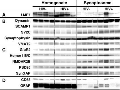
Infection of the central nervous system with human immunodeficiency virus type 1 (HIV-1) can produce morphological changes in the neocortical synaptodendritic arbor that are correlated with neurocognitive impairment. To determine whether HIV-1 infection influences the protein composition of human synapses, a proteomic study of isolated nerve endings was undertaken. Synaptosomes from frontal neocortex were isolated using isopyknic centrifugation from 19 human brain specimens. Purity and enrichment were assessed by measuring pre- and postsynaptic protein markers. Two-dimensional polyacrylamide gel electrophoresis and matrix-assisted laser desorption ionization time-of-flight mass spectrometry was used to screen for proteins differentially expressed in HIV/AIDS. The concentrations of 31 candidate protein spots were potentially abnormal in HIV-infected decedents with HIV encephalitis and/or increased expression of immunoproteasome subunits. Immunoblots showed that the concentration of some of them was related to HIV-1 infection of the brain and immunoproteasome (IPS) induction. Synapsin 1b and stathmin were inversely related to brain HIV-1 load; 14-3-3zeta and 14-4-4epsilon proteins were higher in subjects with HIV-1 loads. Perturbed synaptosome proteins were linked with IPS subunit composition, and 14-3-3zeta was histologically colocalized with IPS subunits in stained neocortical neurons. Proteomics illustrates that certain human proteins within the synaptic compartment are involved with changes in the synaptodendritic arbor and neurocognitive impairment in HIV-1-infected people.
Figures


Similar articles
-
SIV-Mediated Synaptic Dysfunction Is Associated with an Increase in Synapsin Site 1 Phosphorylation and Impaired PP2A Activity.J Neurosci. 2019 Aug 28;39(35):7006-7018. doi: 10.1523/JNEUROSCI.0178-19.2019. Epub 2019 Jul 3. J Neurosci. 2019. PMID: 31270156 Free PMC article.
-
Persistent hijacking of brain proteasomes in HIV-associated dementia.Am J Pathol. 2010 Feb;176(2):893-902. doi: 10.2353/ajpath.2010.090390. Epub 2009 Dec 24. Am J Pathol. 2010. PMID: 20035054 Free PMC article.
-
Proteomic analyses of monocyte-derived macrophages infected with human immunodeficiency virus type 1 primary isolates from Hispanic women with and without cognitive impairment.J Neurovirol. 2009 Jan;15(1):36-50. doi: 10.1080/13550280802385505. Epub 2008 Dec 26. J Neurovirol. 2009. PMID: 19115125 Free PMC article.
-
Proteomic analysis of HIV-infected macrophages.J Neuroimmune Pharmacol. 2011 Mar;6(1):89-106. doi: 10.1007/s11481-010-9253-4. Epub 2010 Dec 14. J Neuroimmune Pharmacol. 2011. PMID: 21153888 Free PMC article. Review.
-
Proteomics and metabolomics of HIV-associated neurocognitive disorders: A systematic review.J Neurochem. 2021 May;157(3):429-449. doi: 10.1111/jnc.15295. Epub 2021 Jan 22. J Neurochem. 2021. PMID: 33421125
Cited by
-
Behavioral and histological assessment of a novel treatment of neuroHIV in humanized mice.Res Sq [Preprint]. 2023 Dec 13:rs.3.rs-3678629. doi: 10.21203/rs.3.rs-3678629/v1. Res Sq. 2023. PMID: 38168407 Free PMC article. Preprint.
-
Neuropsychological, Neurovirological and Neuroimmune Aspects of Abnormal GABAergic Transmission in HIV Infection.J Neuroimmune Pharmacol. 2016 Jun;11(2):279-93. doi: 10.1007/s11481-016-9652-2. Epub 2016 Jan 30. J Neuroimmune Pharmacol. 2016. PMID: 26829944 Free PMC article.
-
14-3-3s are potential biomarkers for HIV-related neurodegeneration.J Neurovirol. 2012 Oct;18(5):341-53. doi: 10.1007/s13365-012-0121-2. Epub 2012 Jul 19. J Neurovirol. 2012. PMID: 22811265 Free PMC article. Review.
-
Diagnostic and prognostic biomarkers for HAND.J Neurovirol. 2019 Oct;25(5):686-701. doi: 10.1007/s13365-018-0705-6. Epub 2019 Jan 3. J Neurovirol. 2019. PMID: 30607890 Free PMC article. Review.
-
Microglia proliferation underlies synaptic dysfunction in the prefrontal cortex: implications for the pathogenesis of HIV-1-associated neurocognitive and affective alterations.J Neurovirol. 2023 Aug;29(4):460-471. doi: 10.1007/s13365-023-01147-x. Epub 2023 May 24. J Neurovirol. 2023. PMID: 37222970 Free PMC article.
References
Publication types
MeSH terms
Substances
Grants and funding
LinkOut - more resources
Full Text Sources
Medical
