. 2009 Sep 15;48(36):8644-55.
doi: 10.1021/bi9009055.

Structure of PqsD, a Pseudomonas quinolone signal biosynthetic enzyme, in complex with anthranilate

Affiliations

Structure of PqsD, a Pseudomonas quinolone signal biosynthetic enzyme, in complex with anthranilate

Asim K Bera et al. Biochemistry. .

Abstract

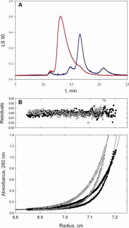
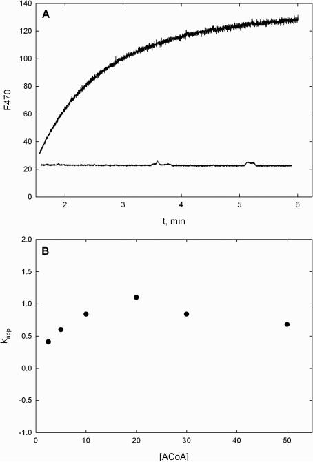
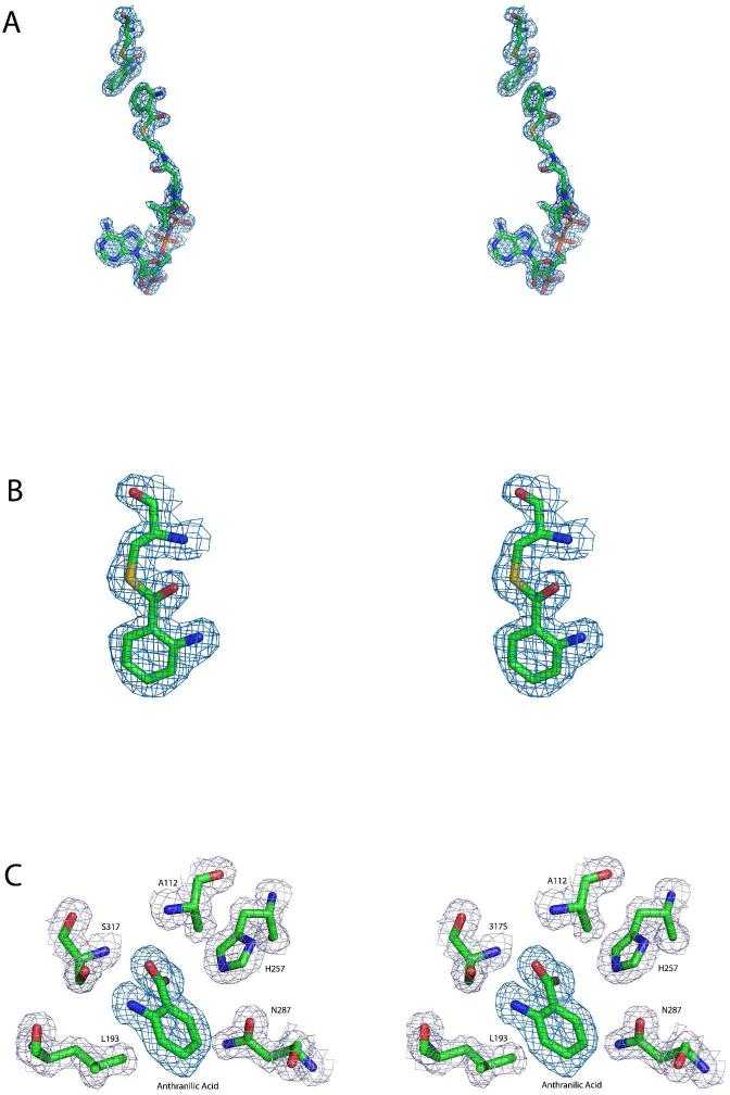
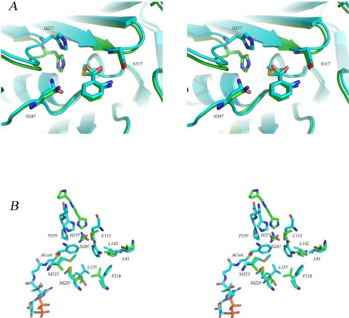
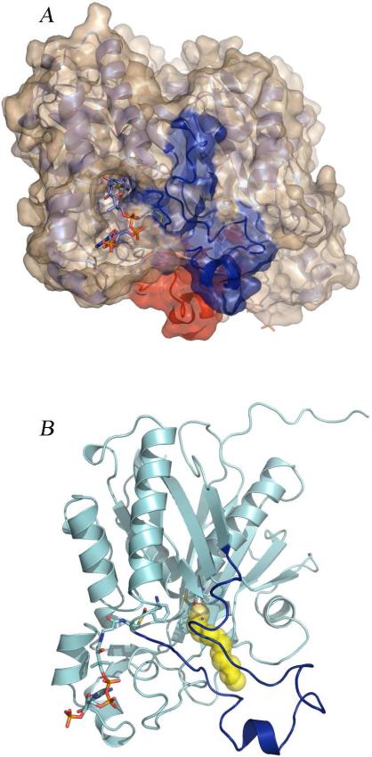
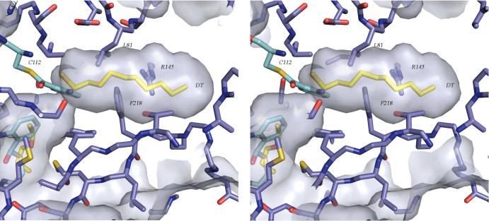
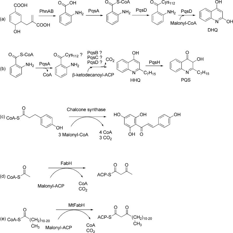
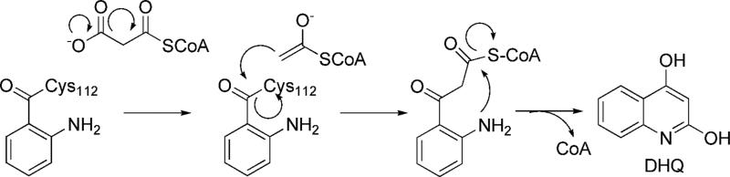
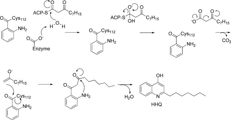
Pseudomonas quinolone signal (PQS), 2-heptyl-3-hydroxy-4-quinolone, is an intercellular alkyl quinolone signaling molecule produced by the opportunistic pathogen Pseudomonas aeruginosa. Alkyl quinolone signaling is an atypical system that, in P. aeruginosa, controls the expression of numerous virulence factors. PQS is synthesized from the tryptophan pathway intermediate, anthranilate, which is derived either from the kynurenine pathway or from an alkyl quinolone specific anthranilate synthase encoded by phnAB. Anthranilate is converted to PQS by the enzymes encoded by the pqsABCDE operon and pqsH. PqsA forms an activated anthraniloyl-CoA thioester that shuttles anthranilate to the PqsD active site where it is transferred to Cys112 of PqsD. In the only biochemically characterized reaction, a condensation then occurs between anthraniloyl-PqsD and malonyl-CoA or malonyl-ACP, a second PqsD substrate, forming 2,4-dihydroxyquinoline (DHQ). The role PqsD plays in the biosynthesis of other alkyl quinolones, such as PQS, is unclear, though it has been reported to be required for their production. No evidence exists that DHQ is a PQS precursor, however. Here we present a structural and biophysical characterization of PqsD that includes several crystal structures of the enzyme, including that of the PqsD-anthranilate covalent intermediate and the inactive Cys112Ala active site mutant in complex with anthranilate. The structure reveals that PqsD is structurally similar to the FabH and chalcone synthase families of fatty acid and polyketide synthases. The crystallographic asymmetric unit contains a PqsD dimer. The PqsD monomer is composed of two nearly identical approximately 170-residue alphabetaalphabetaalpha domains. The structures show anthranilate-liganded Cys112 is positioned deep in the protein interior at the bottom of an approximately 15 A long channel while a second anthraniloyl-CoA molecule is waiting in the cleft leading to the protein surface. Cys112, His257, and Asn287 form the FabH-like catalytic triad of PqsD. The C112A mutant is inactive, although it still reversibly binds anthraniloyl-CoA. The covalent complex between anthranilate and Cys112 clearly illuminates the orientation of key elements of the PqsD catalytic machinery and represents a snapshot of a key point in the catalytic cycle.


Figures

Figure 1
Figure 1
Ribbon diagrams of the PqsD monomer and dimer. In the dimer representation one monomer is shown in cyan and the other is colored by domain with residues 1-174 in blue and 175-329 in red. Anthranilate-modified Cys112 and anthraniloyl-CoA molecules are shown as sticks and illustrate the locations of the active sites and CoA binding tunnels. The PqsD monomer is colored by domain as in the dimer and is rotated slightly to illustrate the structural similarity between the two domains.
Figure 2
Figure 2
Evaluation of the oligomeric state of PqsD. (A) Laser light scattering analysis indicates that PqsD begins to aggregate as it is concentrated. At 1.5 mg/mL (red trace), is predominantly a dimer with a calculated mass of ~72 kDa. At 12 mg/mL (blue trace), PqsD migrates as a species with a calculated mass of greater than 1000 kDa. (B) Analytical ultracentrifugation using sedimentation equilibrium methods confirms that PqsD is a dimer in solution at concentrations examined (0.1-0.5 mg /mL). Representative traces (bottom panel) of absorbance at 280 nm versus radial distance from the center of rotation in centimeters at 15,000 and 19,000 rpm. Distribution of residuals (top panel; Atheor – Aobs) for a single species of molecular weight 72,870.
Figure 3
Figure 3
Evaluation of the interaction between PqsD and ACoA by fluorescence spectroscopy. (A) Changes in fluorescence emission at 470 nm upon mixing 1 μM PqsD or the Cys112Ala mutant with 20 μM ACoA. (B) Plot of kobs vs ACoA using native PqsD revealing that the rate is insensitive to ACoA concentration and is faster than kcat.
Figure 4
Figure 4
The active sites of PqsD and Cys112Ala PqsD are occupied by covalently bound and noncovalently bound ligands. (A) Stereoview of the positive difference density calculated after omitting anthranilate and anthraniloyl-CoA from a round of refinement of the liganded native structure. The map is contoured at 3σ and is depicted along with the ligands from the final refined model. (B) Magnified stereoview of the interaction between Cys112 and anthranilate illustrating the continuous density consistent with a covalent interaction. The displayed omit map was calculated as described for panel A and is shown contoured at 3σ along with the final refined model. (C) Stereoview of the positive difference density, shown in dark blue, calculated after omitting anthranilate from a round of refinement of the Cys112A PqsD structure. A noncovalently bound molecule of anthranilic acid is present in the active site. The map is contoured at 3σ and is depicted along with anthranilate from the final refined model. Nearby side chains are shown along with the final 2Fo-Fc map contoured at 1.2σ.
Figure 5
Figure 5
Comparison of the structure of the PqsD covalent intermediate with the structures of the Cys112Ala mutant and with E. coli FabH. (A) Stereoview of the superimposed active sites of the covalent PqsD-anthranilate complex (blue) and the Cys112Ala mutant structure in complex with anthranilate (green) illustrating the similarity in the positions of the anthraniloyl rings and the differences in the positions of His257. (B) Stereoview of the superimposed active sites of the covalent PqsD-anthranilate complex (blue) and FabH from E. coli (green) illustrating the differences in the position of the active site histidine residue and the possible role of an active site proline. In FabH, His244 is preceded by Pro243 while in PqsD, His257 is followed by Gln258 and Pro259. Also shown is the ACoA molecule occupying the active site entry tunnel and the hydrophobic pocket surrounding the reaction intermediate.
Figure 6
Figure 6
Structural basis for a possible conformational change. (A) The PqsD dimer. Residues 186-222, the regions corresponding to the mobile flap in MtFabH, are colored red in subunit A and blue in subunit B. ACoA is shown in the CoA binding tunnel. (B) Close up of the PqsD monomer illustrating the ‘L’ shaped active site and a possible binding mode for β-ketodecanoate. Residues 186-222 are colored blue. The yellow spheres are decanethiol from the MtFabH structure (PDB code 2QO1). The figure was generated by superimposing PqsD and MtFabH (rmsd = 2.1 Å over 320 residues).
Figure 7
Figure 7
Stereoview of the presumed acyl binding pocket of PqsD illustrating that a conformational change is likely required in order to accommodate β-ketodecanoate. Decanethiol from the MtFabH structure (PDB code 2QO1) was superimposed as in Figure 7 and is shown with yellow carbon atoms. In the observed conformation, Phe218, Leu81, and Arg145 are among the residues limiting access to the binding pocket.
Scheme 1
Scheme 1
Condensation Reactions Catalyzed by PQS Biosynthetic Enzymes (a, b), Chalcone Synthase (c), FabH (d), and MtFabH (e). .
Scheme 2
Scheme 2
Proposed Mechanism for DHQ Formation (13).
Scheme 3
Scheme 3
A Possible Mechanism for HHQ Formation.

References

    1. Wagner VE, Iglewski BH. P. aeruginosa Biofilms in CF Infection. Clin. Rev. Allergy. Immunol. 2008;35:124–134. - PubMed
    1. Parsons JF, Greenhagen BT, Shi K, Calabrese K, Robinson H, Ladner JE. Structural and functional analysis of the pyocyanin biosynthetic protein PhzM from Pseudomonas aeruginosa. Biochemistry. 2007;46:1821–1828. - PMC - PubMed
    1. Engel J, Balachandran P. Role of Pseudomonas aeruginosa type III effectors in disease. Curr. Opin. Microbiol. 2009;12:61–66. - PubMed
    1. Zelenitsky SA, Harding GK, Sun S, Ubhi K, Ariano RE. Treatment and outcome of Pseudomonas aeruginosa bacteraemia: an antibiotic pharmacodynamic analysis. J. Antimicrob. Chemother. 2003;52:668–674. - PubMed
    1. Van Delden C, Iglewski BH. Cell-to-cell signaling and Pseudomonas aeruginosa infections. Emerg. Infect. Dis. 1998;4:551–560. - PMC - PubMed

Publication types

MeSH terms

LinkOut - more resources