

Evaluation of preprocessing steps to compensate for magnetic field distortions due to body movements in BOLD fMRI
- PMID: 19695810
- PMCID: PMC2823819
- DOI: 10.1016/j.mri.2009.07.005
Evaluation of preprocessing steps to compensate for magnetic field distortions due to body movements in BOLD fMRI
Abstract
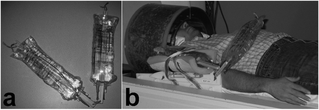
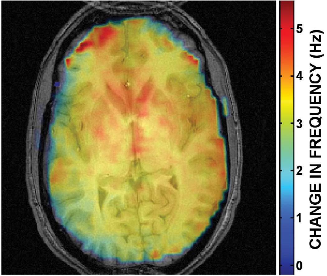
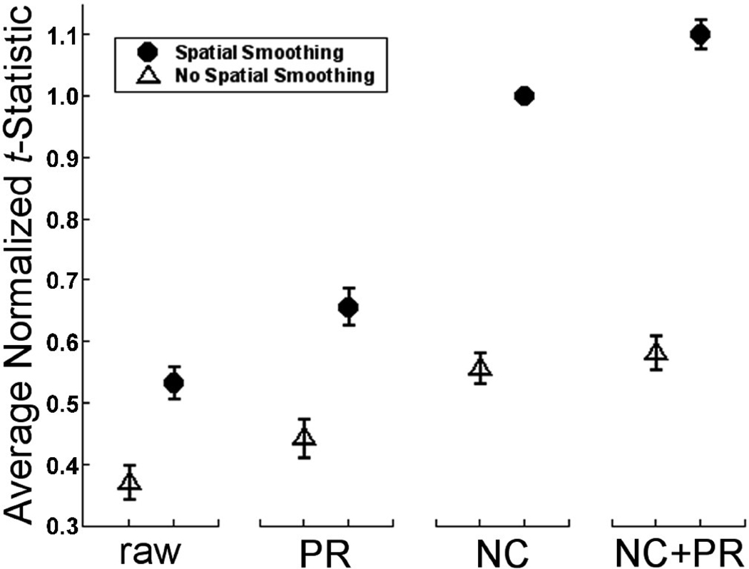
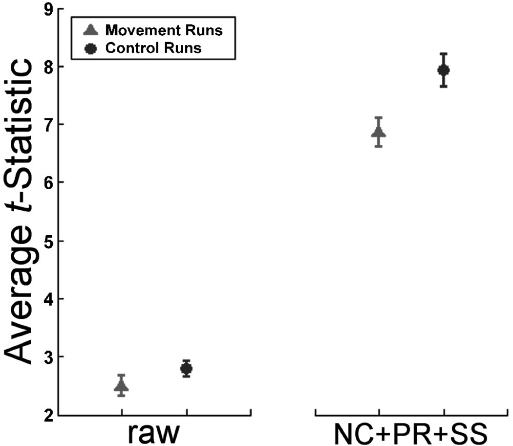
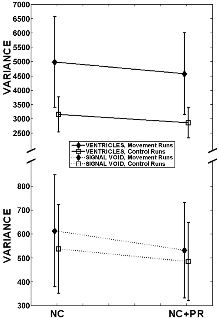
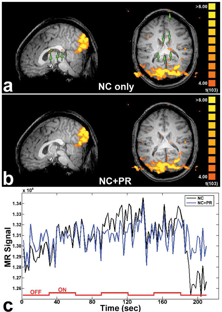
Blood oxygenation level-dependent (BOLD) functional magnetic resonance imaging (fMRI) is currently the dominant technique for non-invasive investigation of brain functions. One of the challenges with BOLD fMRI, particularly at high fields, is compensation for the effects of spatiotemporally varying magnetic field inhomogeneities (DeltaB(0)) caused by normal subject respiration and, in some studies, movement of the subject during the scan to perform tasks related to the functional paradigm. The presence of DeltaB(0) during data acquisition distorts reconstructed images and introduces extraneous fluctuations in the fMRI time series that decrease the BOLD contrast-to-noise ratio. Optimization of the fMRI data-processing pipeline to compensate for geometric distortions is of paramount importance to ensure high quality of fMRI data. To investigate DeltaB(0) caused by subject movement, echo-planar imaging scans were collected with and without concurrent motion of a phantom arm. The phantom arm was constructed and moved by the experimenter to emulate forearm motions while subjects remained still and observed a visual stimulation paradigm. These data were then subjected to eight different combinations of preprocessing steps. The best preprocessing pipeline included navigator correction, a complex phase regressor and spatial smoothing. The synergy between navigator correction and phase regression reduced geometric distortions better than either step in isolation and preconditioned the data to make them more amenable to the benefits of spatial smoothing. The combination of these steps provided a 10% increase in t-statistics compared to only navigator correction and spatial smoothing and reduced the noise and false activations in regions where no legitimate effects would occur.
Copyright 2010 Elsevier Inc. All rights reserved.
Figures
References
-
- Kwong KK, Belliveau JW, Chesler DA, Goldberg IE, Weisskoff RM, Poncelet BP, Kennedy DN, Hoppel BE, Cohen MS, Turner R, Cheng H-M, Brady TJ, Rosen BR. Dynamic magnetic resonance imaging of human brain activity during primary sensory stimulation. Proc Natl Acad Sci USA. 1992;89:5675–5679. - PMC - PubMed
-
- Bandettini PA, Wong EC, Hinks RS, Tikofsky RS, Hyde JS. Time course EPI of human brain function during task activation. Magn Reson Med. 1992;25:390–397. - PubMed
-
- Weisskoff RM, Baker J, Belliveau J, Davis TL, Kwong KK, Cohen MS, Rosen BR. Power spectrum analysis of functional-weighted MR data: what’s in the noise? Proc SMRM. 1993:7.
Publication types
MeSH terms
Grants and funding
LinkOut - more resources
Full Text Sources
Other Literature Sources
Medical
