. 2009 Oct 9;284(41):28367-28381.
doi: 10.1074/jbc.M109.000273. Epub 2009 Aug 19.

Mitotic regulation of the stability of microtubule plus-end tracking protein EB3 by ubiquitin ligase SIAH-1 and Aurora mitotic kinases

Affiliations

Mitotic regulation of the stability of microtubule plus-end tracking protein EB3 by ubiquitin ligase SIAH-1 and Aurora mitotic kinases

Reiko Ban et al. J Biol Chem. .

Abstract

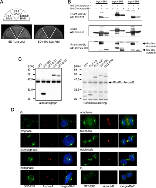
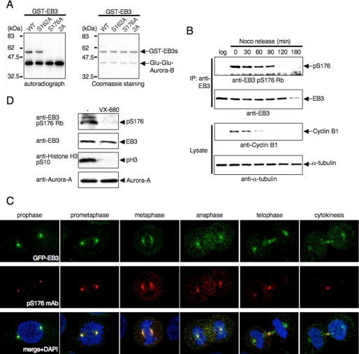
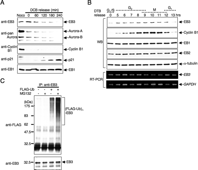
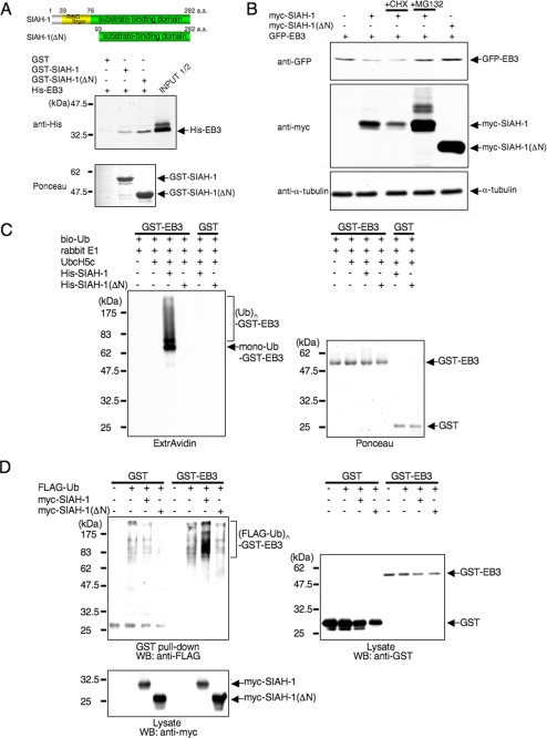
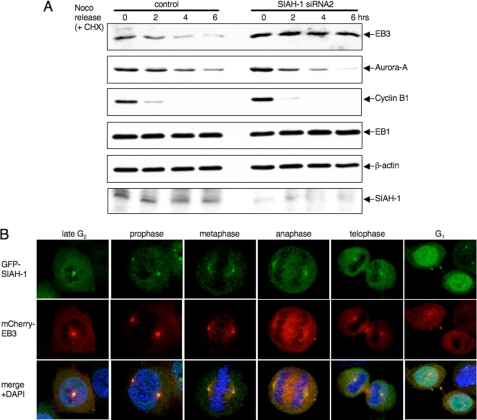
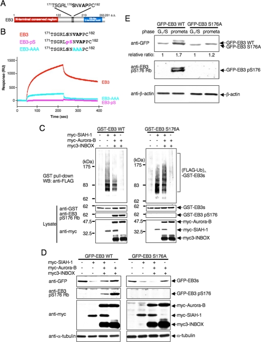
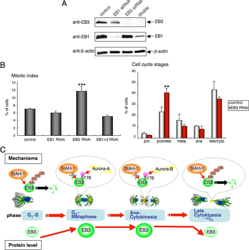
Microtubule plus-end tracking proteins (+TIPs) control microtubule dynamics in fundamental processes such as cell cycle, intracellular transport, and cell motility, but how +TIPs are regulated during mitosis remains largely unclear. Here we show that the endogenous end-binding protein family EB3 is stable during mitosis, facilitates cell cycle progression at prometaphase, and then is down-regulated during the transition to G(1) phase. The ubiquitin-protein isopeptide ligase SIAH-1 facilitates EB3 polyubiquitination and subsequent proteasome-mediated degradation, whereas SIAH-1 knockdown increases EB3 stability and steady-state levels. Two mitotic kinases, Aurora-A and Aurora-B, phosphorylate endogenous EB3 at Ser-176, and the phosphorylation triggers disruption of the EB3-SIAH-1 complex, resulting in EB3 stabilization during mitosis. Our results provide new insight into a regulatory mechanism of +TIPs in cell cycle transition.


Figures

FIGURE 1.
FIGURE 1.
EB2 and EB3, but not EB1, are substrates of Aurora. A, yeast Aurora homolog, Ipl1, binds Bim1, the sole budding yeast member of the human EB1-related family, in a yeast two-hybrid system. Plasmids transformed into PJ69-4A are indicated in each sector. B, binding of EB1 family members and Aurora family members in COS-7 cells that were transfected with the indicated plasmids. The cell lysates were immunoprecipitated (IP) by using anti-Glu-Glu antibody, and the precipitates were analyzed by Western blots (WB) that were probed sequentially with anti-Myc and anti-Glu-Glu antibodies. Asterisk denotes IgG heavy chain. One Western blot shown is representative of three independent experiments. C, in vitro phosphorylation of the EB1 family proteins. GST-fused EB1, EB2, EB3 or H3-(5–15) as a control was incubated with immunoprecipitated Glu-Glu-Aurora-B in the presence of [γ-32P]ATP. The phosphorylation reactions were detected by autoradiography (left panel). The amounts of GST fusion protein used in the assay were compared by Coomassie Blue staining (right panel). A representative result of three independent experiments is shown. D, representative confocal images of EB3 localization during each stage of mitosis by immunofluorescence staining of HeLa cells that stably expressed GFP-EB3. Green, GFP-EB3; red, Aurora-A (left panels) or Aurora-B (right panels); blue, DAPI.
FIGURE 2.
FIGURE 2.
Phosphorylation of EB3 Ser-176 by Aurora-A and Aurora-B during mitosis. A, in vitro phosphorylation of EB3 mutants. The experiment was performed as described in Fig. 1C using GST-fused EB3 mutants and immunoprecipitated Glu-Glu-Aurora-B. The autoradiographs shown are representative of three independent experiments. WT, wild type. B, phosphorylation of endogenous EB3 in mitotic cells. HeLa cells were released from a nocodazole block for the indicated times. Noco and log indicate nocodazole and logarithmic phase, respectively. Immunoprecipitation (IP) was performed with anti-EB3 monoclonal antibody, and the precipitates were analyzed by Western blots probed sequentially with anti-phospho-EB3(Ser-176) rabbit polyclonal antibody (Rb). A representative result of three independent experiments is shown. C, representative confocal images of localization of phospho-EB3(Ser-176) during each stage of mitosis. Immunofluorescence staining of HeLa cells that stably expressed GFP-EB3 was performed using anti-phospho-EB3(Ser-176) mouse monoclonal antibody (mAb). Red, phospho-EB3(Ser-176); green, GFP-EB3; blue, DAPI. D, effects of Aurora inhibitor on EB3 phosphorylation. HeLa cells were arrested at prometaphase with nocodazole and subsequently treated with VX-680 for 4 h, and then the cell lysates were analyzed by Western blots (antibodies indicated at the side of the figure). A representative result of three independent experiments is shown.
FIGURE 3.
FIGURE 3.
EB3 protein level is regulated by the ubiquitin-proteasome system. A, down-regulation of EB3 during the mitosis-to-G1 transition. Lysates from HeLa cells that were released from a DCB block for the indicated times were subjected to Western blots (antibodies indicated on the side of the figure). Noco indicates nocodazole. B, cell cycle-dependent protein expression of EB3. HeLa cells were synchronized at G1/S phase by double thymidine block (DTB) release, and then cells were collected at the indicated times after release from arrest. The cell lysates were used for Western blots (WB) to determine each protein level, and RNAs extracted from each cell were used for reverse transcription-PCR (RT-PCR). C, polyubiquitination of endogenous EB3 in cells. HeLa cells were transfected with plasmids encoding FLAG-ubiquitin. After 24 h, cells were treated with or without 20 μm MG132 for 6 h. Immunoprecipitation (IP) was performed with anti-EB3 antibody, and the precipitates were analyzed by Western blots probed sequentially with anti-FLAG and anti-EB3 antibodies. The results shown are from one representative experiment out of at least three independent experiments.
FIGURE 4.
FIGURE 4.
SIAH-1 is E3 for EB3. A, schematic representation of full-length SIAH-1 and SIAH-1(ΔN) (upper panels) and in vitro binding of EB3 and SIAH-1 (lower panels). Purified His-EB3 was incubated with GST-fused, full-length SIAH-1 and SIAH-1(ΔN) trapped on glutathione-Sepharose beads. The bound EB3 was then eluted and detected by Western blots with anti-penta-His antibody (middle panel). The bottom panel shows Ponceau staining of the GST fusion protein samples used in the assay. a.a., amino acids. B, SIAH-1 targets EB3 for proteasomal degradation. COS-7 cells were transfected with the indicated plasmids. After 48 h, cells were treated with or without 10 μg/ml CHX or 20 μm MG132 for 6 h. Using these cell lysates, Western blots were performed with the indicated antibodies. C, SIAH-1-mediated ubiquitination (Ub) of EB3 in vitro (left panel). The ubiquitination reaction was performed as described under “Experimental Procedures.” The ubiquitinated proteins in the reaction were detected by Western blots using ExtrAvidin. The right panel shows Ponceau staining of the GST fusion protein samples used in the assay. D, SIAH-1-mediated EB3 ubiquitination in cells. COS-7 cells were transfected with the indicated plasmids. After 48 h, cells were treated with 20 μm MG132 for 6 h. GST pulldown was performed, and the precipitates were analyzed by Western blots (WB) probed with anti-FLAG antibody. The results shown are from one representative experiment out of at least three independent experiments.
FIGURE 5.
FIGURE 5.
Degradation of EB3 by SIAH-1 at the mitosis to G1 transition. A, effects of SIAH-1 knockdown on EB3 stability during the mitosis-to-G1 transition. HeLa cells were transfected with siRNA for SIAH-1. After 48 h, nocodazole was added for 16 h; subsequently, cells were released from a nocodazole (Noco) block for the indicated times in the presence of 10 μg/ml CHX. The cell lysates were subjected to Western blots with the indicated antibodies. One Western blot shown is representative of three independent experiments. B, representative confocal images of localization of SIAH-1 and EB3 during each stage of mitosis. Immunofluorescence staining is shown of HeLa cells that stably expressed GFP-SIAH-1 and mCherry-EB3. Green, GFP-SIAH-1; red, mCherry-EB3; blue, DAPI.
FIGURE 6.
FIGURE 6.
Auroras regulate SIAH-1-dependent EB3 degradation through Ser-176 phosphorylation of EB3. A, schematic diagram of a potential SIAH-binding motif (V178AP180) in EB3. Ser-176 is an Aurora phosphorylation site. a.a., amino acids. B, top, schematic diagram of the sequences of wild-type and mutant peptides derived from EB3. Bottom, a BIAcore sensorgram shows that wild-type EB3 peptides, but not mutant EB3 peptides, bind to immobilized SIAH-1. A representative result of four independent experiments is shown. C, effects of Aurora phosphorylation on SIAH-1-mediated ubiquitination of EB3 in cells. COS-7 cells were transfected with each plasmid (as indicated above the figure). The ubiquitinated proteins were detected as described in Fig. 4D. WT, wild type. D, effects of Aurora phosphorylation on SIAH-1-mediated EB3 degradation. COS-7 cells were transfected with each plasmid (as indicated above the figure). After 48 h, cells were harvested, and Western blots (WB) were performed with the indicated antibodies. E, effects of Aurora phosphorylation on the stability of the EB3 proteins in mitosis. HeLa cells that stably expressed each GFP-wild-type EB3 or EB3 S176A were synchronized at each phase. Subsequently, cells were treated with CHX for 6 h, and the cell lysates were subjected to Western blots with the indicated antibodies. The relative protein level of GFP-EB3 at G1/S phase against the prometaphase is shown below each lane. C–E are representative of three independent experiments.
FIGURE 7.
FIGURE 7.
Effects of EB3 knockdown on mitotic progression. A, knockdown of endogenous EB1 and/or EB3. HeLa cells were treated with siRNAs for EB1 and/or EB3 for 72 h, and then the cell lysates were subjected to Western blots with the indicated antibodies. One Western blot shown is representative of three independent experiments. B, left panel, quantitation of the mitotic index of HeLa cells treated with each siRNA after 72 h. The mitotic index is expressed as the percentage of the total population of cells in mitosis (n = 1000). Right panel, quantitation of the cell cycle stages of HeLa cells treated with control or EB3 siRNA after 72 h. Cell cycle stages were determined by scoring all mitotic cells by immunofluorescence for α-tubulin and chromosome staining (n = 1000). C, schematic representation of our results.

References

    1. Steinmetz M. O. (2007) J. Struct. Biol. 158, 137–147 - PubMed
    1. Howard J., Hyman A. A. (2007) Curr. Opin. Cell Biol. 19, 31–35 - PubMed
    1. Cassimeris L. (1999) Curr. Opin. Cell Biol. 11, 134–141 - PubMed
    1. Morrison E. E. (2007) Cell. Mol. Life Sci. 64, 307–317 - PMC - PubMed
    1. Akhmanova A., Steinmetz M. O. (2008) Nat. Rev. Mol. Cell Biol. 9, 309–322 - PubMed

Publication types

MeSH terms