. 2009 Oct 9;284(41):28180-28187.
doi: 10.1074/jbc.M109.036566. Epub 2009 Aug 19.

Akt increases sarcoplasmic reticulum Ca2+ cycling by direct phosphorylation of phospholamban at Thr17

Affiliations

Akt increases sarcoplasmic reticulum Ca2+ cycling by direct phosphorylation of phospholamban at Thr17

Daniele Catalucci et al. J Biol Chem. .

Abstract

Cardiomyocytes adapt to physical stress by increasing their size while maintaining cell function. The serine/threonine kinase Akt plays a critical role in this process of adaptation. We previously reported that transgenic overexpression of an active form of Akt (Akt-E40K) in mice results in increased cardiac contractility and cell size, as well as improved sarcoplasmic reticulum (SR) Ca(2+) handling. Because it is not fully elucidated, we decided to study the molecular mechanism by which Akt-E40K overexpression improves SR Ca(2+) handling. To this end, SR Ca(2+) uptake and the phosphorylation status of phospholamban (PLN) were evaluated in heart extracts from wild-type and Akt-E40K mice and mice harboring inducible and cardiac specific knock-out of phosphatidylinositol-dependent kinase-1, the upstream activator of Akt. Moreover, the effect of Akt was assessed in vitro by overexpressing a mutant Akt targeted preferentially to the SR, and by biochemical assays to evaluate potential interaction with PLN. We found that when activated, Akt interacts with and phosphorylates PLN at Thr(17), the Ca(2+)-calmodulin-dependent kinase IIdelta site, whereas silencing Akt signaling, through the knock-out of phosphatidylinositol-dependent kinase-1, resulted in reduced phosphorylation of PLN at Thr(17). Furthermore, overexpression of SR-targeted Akt in cardiomyocytes improved Ca(2+) handling without affecting cell size. Thus, we describe here a new mechanism whereby the preferential translocation of Akt to the SR is responsible for enhancement of contractility without stimulation of hypertrophy.


Figures

FIGURE 1.
FIGURE 1.
Phosphorylation of PLN at Thr17 is not CAMKIIδ-dependent in Akt-E40K Tg mice. A, left, total extracts from WT and Tg mouse hearts were separated by SDS-PAGE under reducing conditions and incubated with anti-phospho-Thr17-PLN, anti-phospho-Ser16-PLN, anti-total PLN, and anti-actin antibodies. Increased phosphorylation of PLN at Thr17 is evident in the Tg lane. Right, densitometric analysis presented relative to WT (n = 3). *, p < 0.05 Tg versus WT. B, left, blots of cytosolic and SR extracts from WT and Tg mouse hearts were incubated with anti-phospho-CAMKIIδ, anti-total CAMKIIδ, and anti-actin antibodies. Surprisingly, CAMKIIδ phosphorylation is decreased in Tg hearts compared with WT. Right, densitometric analysis presented relative to WT (n = 3). *, p < 0.05 Tg versus WT.
FIGURE 2.
FIGURE 2.
Reduced phosphorylation of PLN at Thr17 in PDK1 knock-out mice is dependent on Akt. Total extracts from WT and PDK1 knock-out (KO) mouse hearts were processed for Western blotting. The results obtained suggest that phosphorylation of PLN at Thr17 is Akt-dependent and not CAMKIIδ-dependent. Right, densitometric analysis (n = 3 for each of the presented data). *, p < 0.05; **, p < 0.01 KO versus WT.
FIGURE 3.
FIGURE 3.
Akt can translocate to the SR where it interacts with PLN. A, blots of cytosolic and SR extracts were incubated with anti-phospho-Akt, anti-HA, and anti-actin antibodies. A phospho-Akt signal was found in both cytosolic and SR extracts from Tg mice, indicating that Akt-E40K localizes also at the SR. B, immunofluorescence staining of cardiomyocytes isolated from WT hearts and labeled with anti-total Akt (green) and anti-PLN (red) antibodies. Akt partially colocalizes with PLN (yellow) after stimulation with insulin, indicating that endogenous Akt can translocate to the SR. Bar represents 4 μm. C, interaction of Akt with PLN. Total lysates from WT and Tg hearts were immunoprecipitated (IP) with anti-HA beads (HA) and Western blots performed with anti-HA and anti-total PLN antibodies. PLN was only detected after immunoprecipitation when extracts from Tg mice were used. For total lysate, one-tenth of the amount used for coimmunoprecipitation experiments was loaded onto gels. D, Akt phosphorylates recombinant PLN in vitro. Recombinant PLN was incubated with or without a commercially available purified active Akt in an in vitro kinase assay. Samples were subsequently separated by SDS-PAGE and probed with anti-phospho-Thr17-PLN, anti-phospho-Ser16-PLN, and total PLN antibodies. A band corresponding to PLN was detected with anti-phospho-Thr17-PLN but not with anti-Ser16-PLN. The blots shown are representative of three independent experiments.
FIGURE 4.
FIGURE 4.
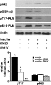
Akt phosphorylates PLN at Thr17 in WT CMs stimulated with insulin. Top, cultured WT CMs were treated with 0.1 μm insulin for 40 min and, where given, pretreated with inhibitors (0.1 μm KN93 or 10 μm AKT inhibitor IV) for 10 min. Representative blot of phosphorylated Akt, GSK3α/β, and PLN at Thr17 and Ser16 is shown. Bottom, densitometric analysis of PLN phosphorylation in insulin-treated cells relative to untreated cells, expressed as mean ± S.D. (n = 3). Statistical significance was assayed by ANOVA. *, p < 0.05; **, p < 0.01 versus insulin-only treated CMs; α, p < 0.05 insulin-only treated versus untreated cells.
FIGURE 5.
FIGURE 5.
SR45Ca2+ uptake in WT and Tg heart homogenate. SR Ca2+ uptake assays were performed at room temperature with homogenates of Tg and WT ventricles at 20 nm free Ca2+ (n = 3). 45Ca2+ uptake (nmol/mg protein/min) was calculated from the slope of the linear regression analysis. The effects of Akt and CAMKII inhibitors (40 μm Akt-IV and 5 μm KN93, respectively) on SR Ca2+ uptake are also given. Values are mean ± S.D. (n = 3). Statistical significance was assayed by ANOVA. *, p < 0.001 versus WT; ζ, p < 0.001 versus untreated Tg.
FIGURE 6.
FIGURE 6.
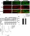
Features of SR-targeted active Akt. WT CMs were mock-transduced (A–C), AdAktE40K-transduced (D–F), or AdAktPLN-transduced (G–I). Dual immunostaining with anti-HA tag (A, D, and G) (green) and anti-PLN (B, E, and H) (red) antibodies was performed and analysis carried out by confocal microscopy. Images were merged (C, F, and I) to determine colocalization (yellow) of transgene HA-tagged Akt with PLN, presumably at the SR. Bar is 10 μm. J, Western blot analysis of mock-, AdAktE40K-, and AdAktPLN-transfected WT CMs. Increased phosphorylation at the CAMKIIδ site, but not at the PKA site, of PLN is evident in CMs transduced with either AdAktE40K or AdAktPLN. Representative blots are shown. K, representative Ca2+ transient traces. L, targeting of active Akt to the SR does not cause CM hypertrophy. Hypertrophy was evaluated as the rate of incorporation of [3H]leucine. Statistical significance was assessed by ANOVA. *, p < 0.05, n = 3.

References

    1. Alessi D. R., Andjelkovic M., Caudwell B., Cron P., Morrice N., Cohen P., Hemmings B. A. (1996) EMBO J. 15, 6541–6551 - PMC - PubMed
    1. Cantley L. C. (2002) Science 296, 1655–1657 - PubMed
    1. Brazil D. P., Hemmings B. A. (2001) Trends Biochem. Sci. 26, 657–664 - PubMed
    1. McMullen J. R., Shioi T., Zhang L., Tarnavski O., Sherwood M. C., Kang P. M., Izumo S. (2003) Proc. Natl. Acad. Sci. U.S.A. 100, 12355–12360 - PMC - PubMed
    1. Condorelli G., Drusco A., Stassi G., Bellacosa A., Roncarati R., Iaccarino G., Russo M. A., Gu Y., Dalton N., Chung C., Latronico M. V., Napoli C., Sadoshima J., Croce C. M., Ross J., Jr. (2002) Proc. Natl. Acad. Sci. U.S.A. 99, 12333–12338 - PMC - PubMed

Publication types

MeSH terms

Substances