. 2009 Oct;37(18):6135-47.
doi: 10.1093/nar/gkp665. Epub 2009 Aug 20.

Differential contribution of the m7G-cap to the 5' end-dependent translation initiation of mammalian mRNAs

Affiliations

Differential contribution of the m7G-cap to the 5' end-dependent translation initiation of mammalian mRNAs

Dmitri E Andreev et al. Nucleic Acids Res. 2009 Oct.

Abstract

Many mammalian mRNAs possess long 5' UTRs with numerous stem-loop structures. For some of them, the presence of Internal Ribosome Entry Sites (IRESes) was suggested to explain their significant activity, especially when cap-dependent translation is compromised. To test this hypothesis, we have compared the translation initiation efficiencies of some cellular 5' UTRs reported to have IRES-activity with those lacking IRES-elements in RNA-transfected cells and cell-free systems. Unlike viral IRESes, the tested 5' UTRs with so-called 'cellular IRESes' demonstrate only background activities when placed in the intercistronic position of dicistronic RNAs. In contrast, they are very active in the monocistronic context and the cap is indispensable for their activities. Surprisingly, in cultured cells or cytoplasmic extracts both the level of stimulation with the cap and the overall translation activity do not correlate with the cumulative energy of the secondary structure of the tested 5' UTRs. The cap positive effect is still observed under profound inhibition of translation with eIF4E-BP1 but its magnitude varies for individual 5' UTRs irrespective of the cumulative energy of their secondary structures. Thus, it is not mandatory to invoke the IRES hypothesis, at least for some mRNAs, to explain their preferential translation when eIF4E is partially inactivated.


Figures

Figure 1.
Figure 1.
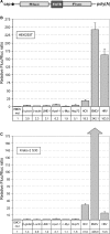
Effect of RNA interference against Rluc in cells transfected by DNA dicistronic constructs on the activity of the second cistron (Fluc) directed by different 5′ UTRs. (A) Schematic presentation of the DNA dicistronic constructs used in the experiments. (B) Results of the DNA transfections of HEK293T cells with and without pRLi plasmid. For each dicistronic construct the activities of Rluc and Fluc in the presence of pRLi were normalized to those in its absence, which were set to 100%. pR+pF is a control test where two monocistronic constructs pR and pF were contransfected with or without pRLi.
Figure 2.
Figure 2.
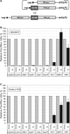
Conventional test to identify IRES elements in the 5′ UTRs of cellular and viral mRNAs using dicistronic RNA constructs. (A) Schematic presentation of the dicistronic RNA construct used to identify IRES elements within 5′ UTRs of cellular mRNAs. The length of poly(A) tail was 50 A residues. (B) Results of transfection of HEK293T cells with dicistronic mRNAs carrying in the intercistronic position various cellular 5′ UTRs or viral IRESes. Fluc/Rluc ratio for the control EMCVmut construct was set to 1. Other Fluc/Rluc ratios were normalized to the Fluc/Rluc ratio of the EMCVmut and, therefore, the values in the bottom row reflect the extent of Fluc stimulation by different 5′ UTRs in the intercistronic position over the EMCVmut control. (C) Translation of the same constructs in the nuclease untreated cytoplasmic extract of ascite carcinoma cells Krebs-2.
Figure 3.
Figure 3.
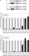
Comparison of activities of various 5′ UTRs in monocistronic versus dicistronic contexts. (A) Schematic representation of mono- and dicistronic mRNAs used in the assay. (B) HEK293T cells were transfected with either dicistronic mRNA derivatives indicated or equimolar mix of the corresponding monocistronic reporter (coding for Fluc) and the normalizing mRNA (Rluc). Fluc/Rluc values for monocistronic mRNAs was taken as 100% for each individual construct. (C) Translation of the same constructs, as in (B), in vitro, in the cytoplasmic S30 extract from cells Krebs-2.
Figure 4.
Figure 4.
Cap-dependence of Fluc mRNAs with various cellular and viral 5′ UTRs. (A) Schematic representation of pairs of monocistronic mRNAs used in the assay. Note: the same reference mRNA, cap-Rluc-poly(A), was used to normalize activities of capped and uncapped (A-capped) Fluc constructs containing the tested 5′ UTRs. To avoid an interference between capped and uncapped transcripts, the cap-Rluc-poly(A) was taken in a very low amount (1 ng) in these experiments. (B) Comparison of indicated capped vs. uncapped monocistronic transcripts in the RNA transfection assay for HEK293T cells. Fluc/Rluc values for capped mRNAs were taken as 100% for each individual construct. (C) Translation of the same pairs of constructs as in (B) and (C) in the S30 extract from cells Krebs-2.
Figure 5.
Figure 5.
Differential inhibition of translation of capped mRNAs containing different cellular 5′ UTRs by m7GTP or 4E-BP1 in the cytoplasmic extract of cells Krebs-2. (A) Interpretative drawing showing distinct mechanisms of inhibition of the cap-dependent translation by m7GTP or eIF4E-BP. (B) Titration curves to determine the concentrations of m7GTP or 4E-BP1 which exert maximum inhibitory effects on the translation of the standard cap-dependent mRNA (β-actin 5′ UTR directed Fluc mRNA). (C) and (D) Effects of m7GTP (100 µM) and 4E-BP1 (0.1 mg/ml) on the translation of m7G-capped and uncapped (A-capped) monocistronic mRNAs containing various cellular 5′ UTRs and viral IRESes in the cytoplasmic extract of cells Krebs-2. As before, the extract was not treated with micrococcal nuclease to ensure competitive conditions for translation of exogenous mRNAs.
Figure 6.
Figure 6.
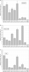
Demonstration of efficient cap-dependent translation initiation of mRNAs with long and highly structured 5′ UTRs both in transfected cells and the nuclease untreated cytoplasmic extract from ascite carcinoma cells Krebs-2. (A) Translation in HEK293T. Exactly the same molar amounts of m7G-capped and polyadenylated mRNAs carrying different 5′ UTRs were used in transfection assays. Viral IRESes were not capped (A-capped) to assess their actual translation initiation potentials in these cell lines. (B) and (C) The same mRNAs but translated in the extract from cells Krebs-2 and RRL, respectively. In all experiments Fluc activities of these constructs were normalized to that for the Fluc mRNA directed by β-globin 5′ UTR.
Figure 7.
Figure 7.
Mutation analysis of the 5′ UTR of Apaf-1 mRNA to reveal the mechanism by which ribosomes select the start codon. (A) Schematic presentation of the secondary structure of the 5′ UTR of Apaf-1 mRNA [adapted from ref. (40)]. The deletions of principal structural domains (mut Δ1 through mut Δ4) are denoted by numbers. Positions of additional upstream start sites (AUG1 and AUG2) are indicated with arrows; natural uAUG triplets are also shown. (B) Fluc expression from transcripts with additional uAUG1 and uAUG2 codons in transfected HEK293T cells. Similar transcripts with uUAGs in analogous positions were used as control; (C) Effect of the deletions shown in (A) on the translation efficiency of the corresponding transcripts, both m7G and A-capped, in HEK293T cells.

References

    1. Pestova TV, Kolupaeva VG, Lomakin IB, Pilipenko EV, Shatsky IN, Agol VI, Hellen CU. Molecular mechanisms of translation initiation in eukaryotes. Proc. Natl Acad. Sci. USA. 2001;98:7029–7036. - PMC - PubMed
    1. Pelletier J, Sonenberg N. Internal initiation of translation of eukaryotic mRNA directed by a sequence derived from poliovirus RNA. Nature. 1988;334:320–325. - PubMed
    1. Pestova TV, Hellen CU, Shatsky IN. Canonical eukaryotic initiation factors determine initiation of translation by internal ribosomal entry. Mol. Cell Biol. 1996;16:6859–6869. - PMC - PubMed
    1. Pestova TV, Shatsky IN, Fletcher SP, Jackson RJ, Hellen CU. A prokaryotic-like mode of cytoplasmic eukaryotic ribosome binding to the initiation codon during internal translation initiation of hepatitis C and classical swine fever virus RNAs. Genes Dev. 1998;12:67–83. - PMC - PubMed
    1. Pilipenko EV, Pestova TV, Kolupaeva VG, Khitrina EV, Poperechnaya AN, Agol VI, Hellen CU. A cell cycle-dependent protein serves as a template-specific translation initiation factor. Genes Dev. 2000;14:2028–2045. - PMC - PubMed

Publication types