Comparative Study
. 2009 Nov;76(5):1082-93.
doi: 10.1124/mol.109.060160. Epub 2009 Aug 20.

Caveolin-1 and lipid microdomains regulate Gs trafficking and attenuate Gs/adenylyl cyclase signaling

Affiliations
Comparative Study

Caveolin-1 and lipid microdomains regulate Gs trafficking and attenuate Gs/adenylyl cyclase signaling

John A Allen et al. Mol Pharmacol. 2009 Nov.

Expression of concern in

Abstract

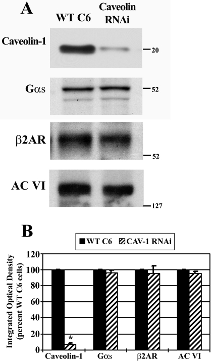
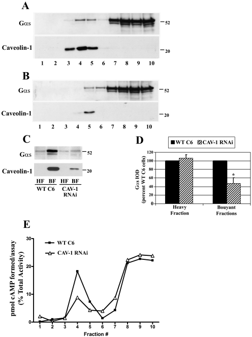
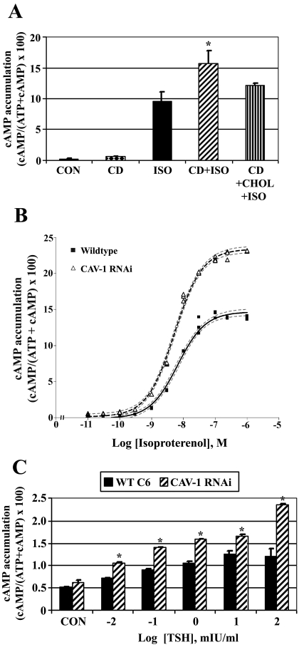
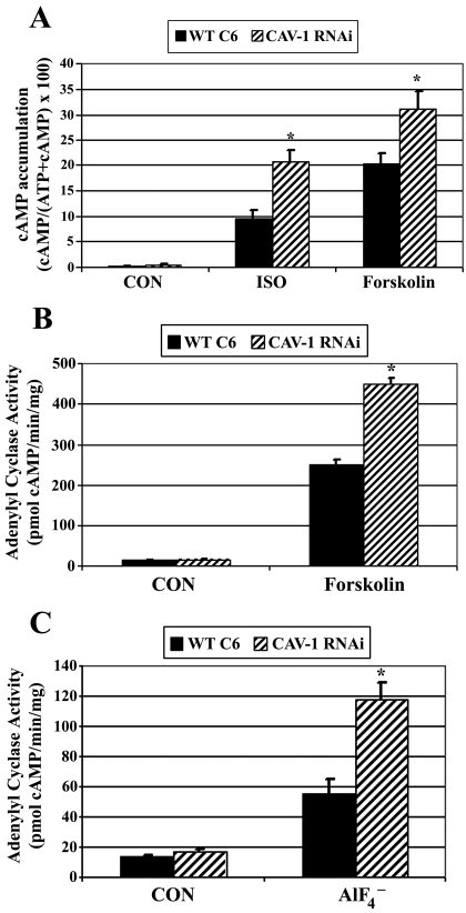
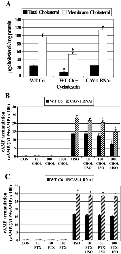
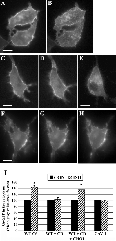
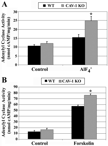
Lipid rafts and caveolae are specialized membrane microdomains implicated in regulating G protein-coupled receptor signaling cascades. Previous studies have suggested that rafts/caveolae may regulate beta-adrenergic receptor/Galpha(s) signaling, but underlying molecular mechanisms are largely undefined. Using a simplified model system in C6 glioma cells, this study disrupts rafts/caveolae using both pharmacological and genetic approaches to test whether caveolin-1 and lipid microdomains regulate G(s) trafficking and signaling. Lipid rafts/caveolae were disrupted in C6 cells by either short-term cholesterol chelation using methyl-beta-cyclodextrin or by stable knockdown of caveolin-1 and -2 by RNA interference. In imaging studies examining Galpha(s)-GFP during signaling, stimulation with the betaAR agonist isoproterenol resulted in internalization of Galpha(s)-GFP; however, this trafficking was blocked by methyl-beta-cyclodextrin or by caveolin knockdown. Caveolin knockdown significantly decreased Galpha(s) localization in detergent insoluble lipid raft/caveolae membrane fractions, suggesting that caveolin localizes a portion of Galpha(s) to these membrane microdomains. Methyl-beta-cyclodextrin or caveolin knockdown significantly increased isoproterenol or thyrotropin-stimulated cAMP accumulation. Furthermore, forskolin- and aluminum tetrafluoride-stimulated adenylyl cyclase activity was significantly increased by caveolin knockdown in cells or in brain membranes obtained from caveolin-1 knockout mice, indicating that caveolin attenuates signaling at the level of Galpha(s)/adenylyl cyclase and distal to GPCRs. Taken together, these results demonstrate that caveolin-1 and lipid microdomains exert a major effect on Galpha(s) trafficking and signaling. It is suggested that lipid rafts/caveolae are sites that remove Galpha(s) from membrane signaling cascades and caveolins might dampen globally Galpha(s)/adenylyl cyclase/cAMP signaling.


Figures

Fig. 1.
Fig. 1.
Stable knockdown of caveolin-1 has no effect on the content of β-adrenergic-related signaling molecules. Total protein lysates from wild-type C6 or stable caveolin-1 knockdown cells were analyzed by SDS/PAGE and immunoblotting to determine relative protein levels of caveolin-1, Gαs, β2AR, and AC type VI. A, immunoreactivity from a representative immunoblot is shown, where the blot was stripped and reprobed sequentially for each protein. B, blots were quantified by scanning densitometry, and data were pooled from four independent experiments and are expressed as a percentage of wild-type C6 cells (n = 4). *, p < 0.05 versus WT C6.
Fig. 2.
Fig. 2.
Caveolin knockdown decreases Gαs localization and adenylyl cyclase activity in lipid rafts/caveolae. Membranes from wild-type C6 or stable caveolin-1 knockdown cells (Cav-1 RNAi) were homogenized, incubated with 1% Triton X-100 followed by sucrose density gradient fractionation and immunoblotting for endogenous Gαs or caveloin-1. A, representative immunoblot of Gαs (top) or the same blot reprobed for caveoin-1 (bottom) in the 10 fractions isolated from wild-type C6 cells where equal volumes were loaded from each fraction. B, representative immunoblots of Gαs (top) or caveolin-1 (bottom) in fractions obtained from stable caveolin knockdown cells where equal volumes were loaded from each fraction. C, buoyant sucrose fractions 3, 4, and 5 enriched in caveolin (BF), as well as the heavy sucrose fraction number 10 (HF) were isolated, and equal protein concentrations from the fractions were loaded and analyzed by immunoblotting for Gαs (top) or caveolin-1 (bottom). D, quantification of Gαs immunoreactivity in the sucrose heavy fraction or buoyant fractions. Blots were quantified by scanning densitometry, and data were pooled from three independent experiments and are expressed as a percentage of wild-type C6 cells (n = 3). *, p < 0.05 versus wild-type C6 cells. E, 50 μl of each isolated fraction was incubated in an adenylyl cyclase reaction mix containing 50 μM forskolin and 32P-ATP. [32P]cAMP was isolated and total picomoles of cAMP formed in each fraction was determined through scintillation counting. These values were converted to a percentage of total aggregate activity of adenylyl cyclase present in each isolated fraction. Data presented are representative of similar results from two independent experiments.
Fig. 3.
Fig. 3.
Disruption of lipid rafts/caveolae with cyclodextrin or caveolin knockdown increases βAR and TSHR-stimulated cAMP accumulation. Wild-type C6 cells were incubated with [2,8-3H]adenine for 18 h to label cellular ATP. A, cells were treated initially with ± 10 mM methyl-β-cyclodextrin (CD) for 30 min to disrupt lipid rafts/caveolae, or with CD followed by CD-cholesterol complexes (CD+CHOL) for 90 min to restore cholesterol to the cells. Intact cells were subsequently treated with 10 μM isoproterenol (ISO) for 30 min and [2,8-3H]cAMP accumulation was determined. Data presented are the means ± S.E.M. from six independent experiments performed in triplicate (n = 6). *, p < 0.05 versus ISO-treated cells. B, wild-type C6 cells or caveolin-1-stable knockdown cells (CAV-1 RNAi) were incubated with [2,8-3H]adenine for 18 h to label cellular ATP. Intact cells were treated with increasing concentrations of ISO for 30 min and [2,8-3H]cAMP accumulation was determined. Data are the means ± S.E.M. from a single dose-response experiment performed in triplicate with similar results observed in three independent experiments. C, wild-type C6 cells or CAV-1 RNAi cells were transiently transfected with human thyrotropin (TSH) receptor and 24 h later were incubated with [2,8-3H]adenine for 18 h to radioactively label cellular ATP. Intact cells were treated with increasing concentrations of TSH for 30 min, and [2,8-3H]cAMP accumulation was determined. Data are the means ± S.E.M. from three independent experiments. *, p < 0.05 versus WT C6 cells for each treatment group.
Fig. 4.
Fig. 4.
Caveolin knockdown increases forskolin- and fluoride-stimulated Gαs/adenylyl cyclase signaling. A, wild-type C6 cells (WT C6) or caveolin-1 stable knockdown cells (CAV-1 RNAi) were incubated with [2,8-3H]adenine for 18 h to label cellular ATP. Intact cells were treated with ± 10 μM isoproterenol (ISO) or 100 μM forskolin for 30 min, and [2,8-3H]cAMP accumulation was determined. Data presented are the means ± S.E.M. from six independent experiments performed in triplicate (n = 6). *, p < 0.05 versus WT C6 cells for each treatment group. B, adenylyl cyclase activity was determined in cell membranes isolated from WT C6 or CAV-1 RNAi cells. Membranes were incubated with ± 50 μM forskolin for 20 min [32P]cAMP was isolated and determined by scintillation counting, normalized to time and milligrams of membrane protein. Data presented are the means ± S.E.M. from a single experiment performed in triplicate and are representative of similar results observed in three independent experiments. C, adenylyl cyclase activity was determined in cell membranes isolated from WT C6 or CAV-1 RNAi cells. Membranes were incubated for 20 min with a reaction mixture including 32P-ATP and ± 10 mM sodium fluoride/20 μM aluminum chloride. [32P]cAMP was isolated and determined by scintillation counting, normalized to time and milligrams of membrane protein. Data presented are the means ± S.E.M. from an experiment performed in triplicate and is representative of similar results observed in four independent experiments.
Fig. 5.
Fig. 5.
Increased cAMP accumulation due to caveolin knockdown is independent of cholesterol and Gαi. A, analysis of cholesterol content in wild-type C6 cells (WT C6) or caveolin-1 stable knockdown cells (CAV-1 RNAi). WT C6 cells were treated with ± 10 mM methyl-β-cyclodextrin for 30 min. Total cell lysates or membranes from cells were analyzed for cholesterol content. Data presented are the means ± S.E.M. from three independent experiments (n = 3). a, p < 0.05 versus total cholesterol in WT C6 cells; b, p < 0.05 versus membrane cholesterol in WT C6 cells. B, effects of adding exogenous cholesterol on isoproterenol-stimulated cAMP accumulation. Cells were incubated with [2,8-3H]adenine for 18 h to label cellular ATP and subsequently treated with ±10, 100, or 1000 μg/ml cholesterol (CHOL) for 2 h. Intact cells were subsequently treated with ± 10 μM isoproterenol (ISO) for 30 min and [2,8-3H]cAMP accumulation was determined. Data presented are the means ± S.E.M. from three independent experiments performed in triplicate. a, p < 0.05 versus ISO treated CAV-1 RNAi cells; b, p < 0.05 versus ISO-treated WT C6 cells. C, increased cAMP accumulation as a result of caveolin knockdown is independent of Gαi coupling. Cells were incubated with [2,8-3H]adenine for 18 h to label cellular ATP. During this incubation, cells were also treated with 10, 50, or 100 ng/ml pertussis toxin (PTX), which prevents Gαi subunits from coupling to the βAR. Intact cells were subsequently exposed to ±10 μM ISO for 30 min and [2,8-3H]cAMP accumulation was determined. Data presented are the means ± S.E.M. from three independent experiments. *, p < 0.05 versus WT C6 cells in each treatment group.
Fig. 6.
Fig. 6.
Agonist-induced Gαs internalization requires lipid microdomains and caveolin-1. C6 glioma cells were transiently transfected with Gαs-GFP, and trafficking was assessed using digital fluorescence microscopy. Living cells were imaged in real time, and individual frames are shown before and after 10 μM isoproterenol (ISO) treatment. A and B, Gαs-GFP localization in wild-type C6 cells before agonist treatment (A) or after 20 min of isoproterenol stimulation (B). C to E, wild-type C6 cells expressing Gαs-GFP were preincubated with 10 mM methyl-β-cyclodextrin (CD) for 30 min and imaged (C), and cells were subsequently treated with isoproterenol for 30 min (D). These cells were washed and incubated for 90 min with cyclodextrin-cholesterol complexes (CHOL) to restore cellular cholesterol and then were treated again with isoproterenol for 30 min (E). F to H, C6 cells in which caveolin-1 was stably knocked-down by RNAi were transfected with Gαs-GFP and imaged before agonist exposure (F) or after isoproterenol treatment at 15 min (G) and 30 min (H). I, Gαs-GFP internalization in response to agonist was quantified by determining the mean fluorescence intensity in the cytoplasm of cells and is expressed as a percentage control. More than 50 cells in each treatment group were quantified from six or more independent experiments (n = 6). *, p < 0.05 versus CON. Scale bars, 10 μm (all panels).
Fig. 7.
Fig. 7.
Caveolin-1 knockout increases fluoride and forskolin-stimulated adenylyl cyclase activity in mouse striatum. Ten- to 12-week-old wild-type or caveolin-1 knockout littermate mice were colony bred, brains were obtained, and striatum was microdissected. 10 μg of isolated striatum membranes were assayed for adenylyl cyclase activity. A, striatum membranes from wild-type or caveolin-1 knockout mice were incubated for 20 min, with a reaction mixture including [32P]ATP and ±10 mM sodium fluoride/20 μM aluminum chloride. [32P]cAMP was isolated and determined by scintillation counting, normalized to time and milligrams of membrane protein. B, striatum membranes were incubated with ± 100 μM forskolin for 20 min [32P]cAMP was isolated and determined by scintillation counting, normalized to time and milligrams of membrane protein. Data are the means ± S.E.M. from four littermate mice pairs (n = 4) assayed in triplicate. *, p < 0.05 versus wild-type mice for each treatment group.

Similar articles

Cited by

References

    1. Allen JA, Halverson-Tamboli RA, Rasenick MM. (2007) Lipid raft microdomains and neurotransmitter signalling. Nat Rev Neurosci 8:128–140 - PubMed
    1. Allen JA, Yu JZ, Donati RJ, Rasenick MM. (2005) β-Adrenergic receptor stimulation promotes Gαs internalization through lipid rafts: a study in living cells. Mol Pharmacol 67:1493–1504 - PubMed
    1. Bhatnagar A, Sheffler DJ, Kroeze WK, Compton-Toth B, Roth BL. (2004) Caveolin-1 interacts with 5-HT2A serotonin receptors and profoundly modulates the signaling of selected Gαq-coupled protein receptors. J Biol Chem 279:34614–34623 - PubMed
    1. Bradford MM. (1976) A rapid and sensitive method for the quantitation of microgram quantities of protein utilizing the principle of protein-dye binding. Anal Biochem 72:248–254 - PubMed
    1. Brown DA. (2006) Lipid rafts, detergent-resistant membranes, and raft targeting signals. Physiology (Bethesda) 21:430–439 - PubMed

Publication types

MeSH terms

LinkOut - more resources