. 2009 Sep 15;23(18):2224-36.
doi: 10.1101/gad.1844309. Epub 2009 Aug 20.

Releasing cohesin from chromosome arms in early mitosis: opposing actions of Wapl-Pds5 and Sgo1

Affiliations

Releasing cohesin from chromosome arms in early mitosis: opposing actions of Wapl-Pds5 and Sgo1

Keishi Shintomi et al. Genes Dev. .

Abstract

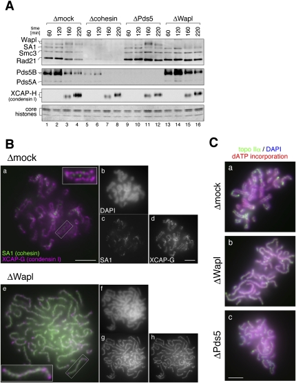
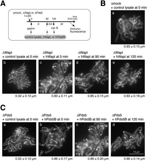
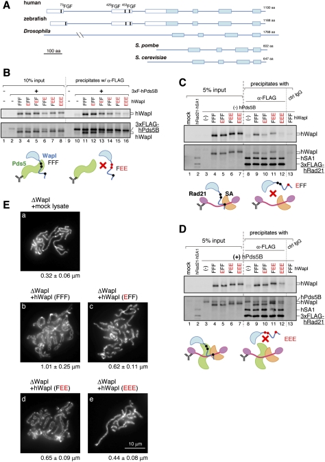
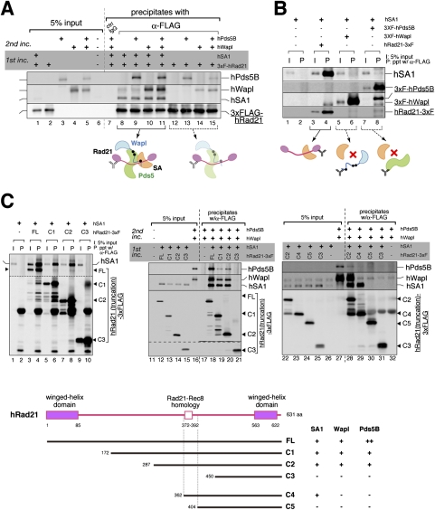
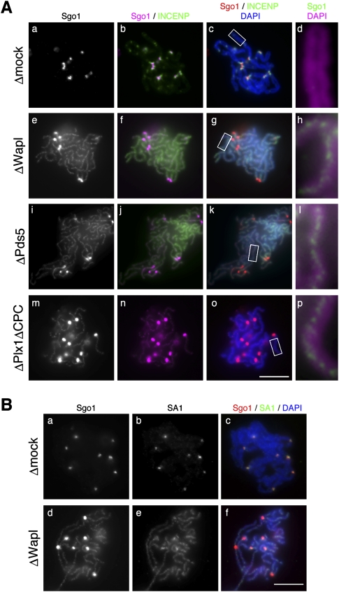
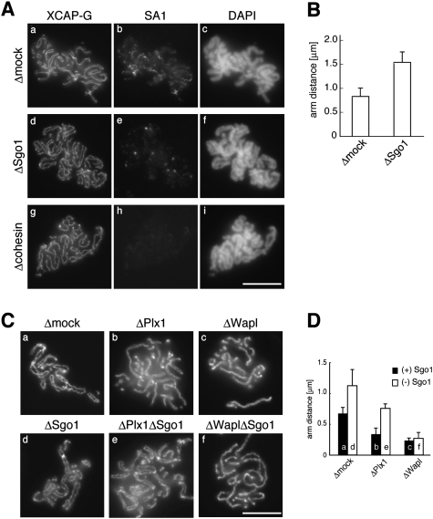
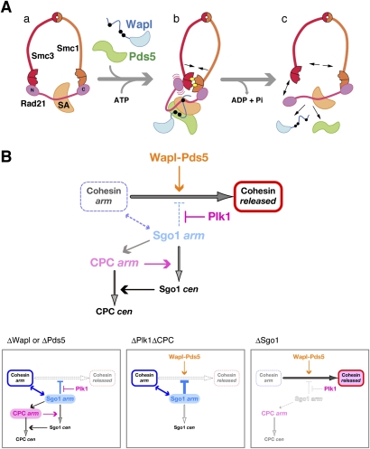
The cohesin complex establishes sister chromatid cohesion during S phase. In metazoan cells, most if not all cohesin dissociates from chromatin during mitotic prophase, leading to the formation of metaphase chromosomes with two cytologically discernible chromatids. This process, known as sister chromatid resolution, is believed to be a prerequisite for synchronous separation of sister chromatids in subsequent anaphase. To dissect this process at a mechanistic level, we set up an in vitro system. Sister chromatid resolution is severely impaired upon depletion of Wapl from Xenopus egg extracts. Exogenously added human Wapl can rescue these defects and, remarkably, it can do so in a very short time window of early mitosis. A similar set of observations is made for Pds5, a factor implicated previously in the stabilization of interphase cohesion. Characteristic amino acid motifs (the FGF motifs) in Wapl coordinate its physical and functional interactions with Pds5 and cohesin subunits. We propose that Wapl and Pds5 directly modulate conformational changes of cohesin to make it competent for dissociation from chromatin during prophase. Evidence is also presented that Sgo1 plays a hitherto underappreciated role in stabilizing cohesin along chromosome arms, which is antagonized by the mitotic kinases polo-like kinsase (Plk1) and aurora B.


Figures

Figure 1.
Figure 1.
Wapl and Pds5 are required for sister chromatid resolution in Xenopus egg extracts. (A) Sperm chromatin was incubated for 120 min with interphase egg extracts that had been depleted with control IgG (Δmock; lanes 1-4), a mixture of anti-Smc1 and anti-Smc3 (Δcohesin; lanes 5-8), a mixture of anti-Pds5A and anti-Pds5B (ΔPds5; lanes 9-12), or anti-Wapl (ΔWapl; lanes 13-16). Cyclin B was added to trigger entry into mitosis, and the mixtures were incubated for another 100 min. At the indicated time points, chromatin-bound proteins were isolated and analyzed by immunoblotting with the antibodies indicated. The lowest part of the gel was stained with Coomassie brilliant blue to determine the level of core histones as a control for chromatin recovery. The efficiency of depletion in each extract is shown in Supplemental Figure S2. (B) Metaphase chromosomes were assembled in mock-depleted (Δmock; panels a–d) or Wapl-depleted (ΔWapl; panels e–h) extracts, fixed, and double-stained with anti-SA1 (green, panels a,e; grayscale images, panels c,g) and anti-XCAP-G (magenta, panels a,e; grayscale images, panels d,h). The insets show close-ups of selected chromosomal regions (indicated by the rectangles). (Panels b,f) Bulk chromosomal DNA was counterstained with DAPI (grayscale images). Bars, 5 μm. (C) Metaphase chromosomes were assembled in mock-depleted (Δmock; panel a), Wapl-depleted (ΔWapl; panel b), or Pds5-depleted (ΔPds5; panel c) extracts that had been supplemented with biotin-dATP. The chromosomes were fixed and stained with anti-topo IIα (green). Bulk and replicated DNA were visualized with DAPI (blue) and FITC-conjugated avidin (red), respectively. Bar, 5 μm.
Figure 2.
Figure 2.
Wapl and Pds5 promote sister chromatid resolution in a short time window upon mitotic entry. (A) Sperm chromatin was added to Xenopus egg interphase extracts that had been immunodepleted with control IgG (Δmock), anti-Wapl (ΔWapl), or anti-Pds5 (ΔPds5). Cyclin B (cyc B) was added at 120 min to drive the cell cycle state into mitosis, and chromosomes were fixed at 210 min for immunofluorescence. A control reticulocyte lysate or a lysate containing hWapl (or hPds5B) was added to the extracts at different time points. (B) Metaphase chromosomes were assembled in mock-depleted (panel a) or Wapl-depleted (panels b–e) extracts that had been supplemented with reticulocyte lysates containing no hWapl (panels a,b) or hWapl (panels c–e) at the indicated time points. The chromosomes were stained with anti-topo IIα as described in Figure 1C. Measurements of the distance between sister chromatid axes were provided below each panel. In all chromosomes shown here, duplication of sister DNA molecules was confirmed as judged by dATP incorporation (data not shown). (C) Metaphase chromosomes were assembled in Pds5-depleted extracts that had been supplemented with reticulocyte lysates containing no Pds5 (panel a) or hPds5B (panels b–d) at the indicated time points. The chromosomes were stained and analyzed as above.
Figure 3.
Figure 3.
The FGF motifs in Wapl play crucial roles in sister chromatid resolution by coordinating its interaction with Pds5 and cohesin. (A) Schematic representation of the primary structure of Wapl orthologs. The filled boxes indicate regions highly conserved from yeasts to humans, whereas the open boxes indicate sequences unique to vertebrate members. Three FGF motifs (thick vertical lines) are found in all of the vertebrate orthologs investigated (see Supplemental Fig. S3). (B) A panel of mutant hWapl was translated in vitro simultaneously with 3xFlag-hPds5B, and then precipitated with anti-Flag beads. The precipitates (lanes 9–16) along with input fractions (lanes 1–8) were analyzed by immunoblotting. Representative results are depicted in the cartoons. (C) A mixture of in vitro translated 3xFlag-hRad21 and hSA1 was precipitated with anti-Flag beads. The beads were then subjected to another incubation with lysates, producing a panel of mutant hWapl. The precipitates (lanes 8–13) and input fractions (lanes 1–7) were analyzed by immunoblotting. (D) The same set of experiments as C was performed, except that hPds5 was coproduced in lysates used for the second incubation. From BD, the membranes were probed first with anti-Wapl (top) and subsequently with a mixture of the antibodies indicated (bottom). (E) Metaphase chromosomes were assembled in a Wapl-depleted extract that had been supplemented with the hWapl mutants indicated. The chromosomes were analyzed as described in Figure 2.
Figure 4.
Figure 4.
The major target of SA1, Wapl, and Pds5 is the central region of Rad21. (A) 3xFlag-hRad21 was translated in vitro individually or simultaneously with hSA1, and then precipitated with anti-Flag beads. The beads were subjected to another incubation with hWapl and hPds5B, which had been translated in various combinations as indicated. After the second incubation, the precipitates (lanes 7–15) and input fractions (lanes 1–6) were analyzed by immunoblotting. (B) hSA1 was translated in vitro alone (lanes 1,2) or simultaneously with 3xFlag-tagged hRad21 (lanes 3,4), 3xFlag-tagged hWapl (lanes 5,6), or 3xFlag-tagged hPds5B (lanes 7,8), and was subjected to immunoprecipitation with anti-Flag. Aliquots of input fractions (lanes 1,3,5,7) and precipitates (lanes 2,4,6,8) were analyzed by immunoblotting. (C) Full-length hRad21-3xFlag (FL) or its truncated versions (C1, C2, C3, C4, and C5) were translated with hSA1 and then precipitated with anti-Flag beads. (Left panel) Five percent of the input fraction (I) and the bead-bound fractions (P) was analyzed by immunoblotting. (Middle and right panels) Alternatively, the beads were subjected to another incubation with hPds5B and hWapl before being analyzed by immunoblotting. A summary of the results is shown in the bottom panel. For sequence alignment of Rad21 and Rec8 in vertebrates, see Supplemental Figure S5. We notice that, compared with hSA1 or hWapl, hPds5B was more sensitive to any truncations of hRad21. Thus, hPds5B may make additional contacts with hRad21 outside of its central region.
Figure 5.
Figure 5.
Sgo1 localizes to the intersister regions in poorly resolved chromosomes. (A) Metaphase chromosomes were assembled in the extracts that had been depleted with control IgG (Δmock; panels a–d), anti-Wapl (ΔWapl; panels e–h), anti-Pds5 (ΔPds5; panels i–l), or a mixture of anti-Plx1, anti-aurora B, and anti-INCENP (ΔPlx1ΔCPC; panels m–p). The chromosomes were fixed and double-stained with anti-Sgo1 (grayscale images, panels a,e,i,m; magenta, panels b,f,j,n; red, panels c,g,k,o; green, panels d,h,l,p) and anti-INCENP (green, panels b,c,f,g,j,k,n,o). DNA was counterstained with DAPI (blue, panels c,g,k,o; magenta, panels d,h,l,p). Close-ups of selected regions of chromosomal arms (indicated by the rectangles in panels c,g,k,o, respectively) are shown in panels d, h, l, and p. (B) Chromosomes that had been assembled in mock-depleted and Wapl-depleted extracts were double-stained with anti-Sgo1 (grayscale, panels a,d; red, panels c,f) and anti-SA1 (grayscale, panels b,e; green, panels c,f). (Panels c,f) DNA was counterstained with DAPI (blue). Bars, 10 μm. The efficiency of depletion in each extract is shown in Supplemental Figure S2.
Figure 6.
Figure 6.
Sgo1 plays a role in stabilizing arm cohesion. (A) Metaphase chromosomes were assembled in a control extract (Δmock; panels a–c), Sgo1-depleted extract (ΔSgo1; panels d–f), or cohesin-depleted extract (Δcohesin; panels g–i), and double-stained with anti-XCAP-G (panels a,d,g) and anti-SA1 (panels b,e,h). (Panels c,f,i) The bulk DNA was counterstained with DAPI. (B) The distance between sister chromatid axes was measured for >50 chromosomes from each condition and plotted. (C) Replicated chromosomes were assembled in a control extract (panel a) or extracts depleted of various factors (individually or in combinations) as indicated (panels b–f), and stained with anti-topo IIα. (D) The distance between sister chromatid axes was measured and plotted as in B. Bars, 10 μm. The efficiency of depletion in each extract is shown in Supplemental Figure S2.
Figure 7.
Figure 7.
Opposing actions of Wapl–Pds5 and Sgo1 on releasing cohesin from chromosome arms. (A) A speculative model for the action of Wapl and Pds5 on cohesin release. (Panels a,b) Wapl and Pds5 interact with the central region of Rad21 to induce its conformational changes upon entry into mitosis, which may in turn facilitate ATP-mediated engagement of the head domains of Smc1 and Smc3. (Panel c) The engaged configuration could transiently destabilize either one of the two SMC–kleisin interfaces, and subsequent ATP hydrolysis allows opening of the cohesin ring, thereby allowing its dissociation from chromatin. ATP molecules are shown by the yellow ovals. (B, top panel) Schematic presentation of a regulatory network for cohesin release from chromosome arms during early mitosis. The two populations of Sgo1 (and the CPC) along chromosome arms and at centromeres are indicated by “arm” and “cen,” respectively. (Bottom panels) This model nicely predicts the behavior of cohesin and its regulators observed under different conditions. See the text for details.

References

    1. Anderson DE, Losada A, Erickson HP, Hirano T. Condensin and cohesin display different arm conformations with characteristic hinge angles. J Cell Biol. 2002;156:419–424. - PMC - PubMed
    1. Bayliss R, Littlewood T, Stewart M. Structural basis for the interaction between FxFG nucleoporin repeats and importin-β in nuclear trafficking. Cell. 2000;102:99–108. - PubMed
    1. Belmont AS, Sedat JW, Agard DA. A three-dimensional approach to mitotic chromosome structure: Evidence for a complex hierarchical organization. J Cell Biol. 1987;105:77–92. - PMC - PubMed
    1. Ben-Shahar TR, Heeger S, Lehane C, East P, Flynn H, Skehel M, Uhlmann F. Eco1-dependent cohesin acetylation during establishment of sister chromatid cohesion. Science. 2008;321:563–566. - PubMed
    1. Bernard P, Schmidt CK, Vaur S, Dheur S, Drogat J, Genier S, Ekwall K, Uhlmann F, Javerzat JP. Cell-cycle regulation of cohesin stability along fission yeast chromosomes. EMBO J. 2008;27:111–121. - PMC - PubMed

Publication types

MeSH terms