Review
. 2009 Aug 21;325(5943):960-3.
doi: 10.1126/science.1168823.

Structural plasticity in actin and tubulin polymer dynamics

Affiliations
Review

Structural plasticity in actin and tubulin polymer dynamics

Hao Yuan Kueh et al. Science. .

Abstract

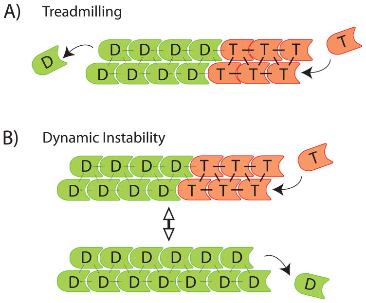
Actin filaments and microtubules polymerize and depolymerize by adding and removing subunits at polymer ends, and these dynamics drive cytoplasmic organization, cell division, and cell motility. Since Wegner proposed the treadmilling theory for actin in 1976, it has largely been assumed that the chemical state of the bound nucleotide determines the rates of subunit addition and removal. This chemical kinetics view is difficult to reconcile with observations revealing multiple structural states of the polymer that influence polymerization dynamics but that are not strictly coupled to the bound nucleotide state. We refer to these phenomena as "structural plasticity" and discuss emerging evidence that they play a central role in polymer dynamics and function.


Figures

Figure 1
Figure 1
Classical chemical kinetics models of polymerization dynamics. T – NTP-bound subunit (red); D – NDP-bound subunit (green). A) Treadmilling. Arrows indicate NTP-subunit association (T, right), and NDP-subunit dissociation from the opposite end (D, left). B) Dynamic instability. Arrows at polymer ends indicate NTP-subunit association (top) and NDP-subunit dissociation (bottom). Bidirectional arrow indicates reversible transitions of the polymer between growing and shrinking states.
Figure 2
Figure 2
Microtubule structural plasticity. A) Cryo-EM images of a growing microtubule end showing a curved, open sheet. B) Cryo-EM image of a shrinking microtubule end showing outwards curled protofilaments. C,D) Localization of microtubule segments with a stable lattice structure, recognized by a recombinant antibody. C. Pure tubulin microtubules (green) growing from a centrosome stained with an antibody that recognizes a stable structural state of the microtubule lattice (red). Note staining of growing tips (white arrows). D) Microtubules in a cell (green) stained with the antibody (red). Note tip staining, presumably on growing microtubles (white arrowhead), lack of tip staining, presumably on shrinking microtubules (empty arrowhead) and internal segments recognized by the antibody (empty arrows). A,B courtesy of T. Hyman, MPI Dresden. C, D adapted from (26).
Figure 3
Figure 3
Actin filament structural plasticity. A) Negative-stain EM images of pure actin filaments two minutes after initiation of polymerization. Note ragged appearance. A′) 3D reconstruction of Cryo-EM images of actin filaments, revealing a structural state in which subunits (numbered) are tilted ~30o relative to the canonical helix. This state is enriched in newly-polymerized filaments. B) EM image of actin filaments aged for 2 hrs. Note regular appearance. B′) 3D reconstruction of Cryo-EM images of actin filaments, revealing a canonical helical state. This canonical state is enriched in aged filaments. Scale bar = 100 nm in both EM images. (Images courtesy of A. Orlova, and E. Egelman, University of Virginia. Reconstructions courtesy of V. Galkin, A. Orlova and E. Egelman, University of Virginia).- again permissions required C) Length vs. time trace for a single fluorescently-labeled actin filament depolymerizing in buffer. Adapted from (36), © 2008, National Academy of Sciences USA. Filament switches from fast to slow depolymerization, and then back to fast. We propose that these switches are caused by spontaneous structural transitions possibly between those shown in A, B.

References

    1. Inoue S, Sato H. J Gen Physiol. 1967 Jul;50(Suppl):259. - PMC - PubMed
    1. Pollard TD, Borisy GG. Cell. 2003 Feb 21;112:453. - PubMed
    1. Mitchison TJ, Salmon ED. Nat Cell Biol. 2001 Jan;3:E17. - PubMed
    1. Dye NA, Shapiro L. Trends Cell Biol. 2007 May;17:239. - PubMed
    1. Egelman EH. Curr Opin Struct Biol. 2003 Apr;13:244. - PubMed

Publication types

MeSH terms