. 2009 Aug 21;325(5943):1014-7.
doi: 10.1126/science.1175275.

Structures of the ribosome in intermediate states of ratcheting

Affiliations

Structures of the ribosome in intermediate states of ratcheting

Wen Zhang et al. Science. .

Abstract

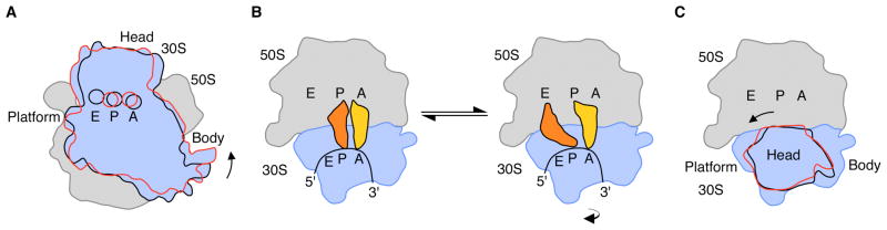
Protein biosynthesis on the ribosome requires repeated cycles of ratcheting, which couples rotation of the two ribosomal subunits with respect to each other, and swiveling of the head domain of the small subunit. However, the molecular basis for how the two ribosomal subunits rearrange contacts with each other during ratcheting while remaining stably associated is not known. Here, we describe x-ray crystal structures of the intact Escherichia coli ribosome, either in the apo-form (3.5 angstrom resolution) or with one (4.0 angstrom resolution) or two (4.0 angstrom resolution) anticodon stem-loop tRNA mimics bound, that reveal intermediate states of intersubunit rotation. In the structures, the interface between the small and large ribosomal subunits rearranges in discrete steps along the ratcheting pathway. Positioning of the head domain of the small subunit is controlled by interactions with the large subunit and with the tRNA bound in the peptidyl-tRNA site. The intermediates observed here provide insight into how tRNAs move into the hybrid state of binding that precedes the final steps of mRNA and tRNA translocation.


Figures

Fig. 1
Fig. 1
Rotated states of the ribosome. (A) View of the bacterial 70S ribosome, composed of the small (30S) ribosomal subunit and the large (50S) ribosomal subunit. The small subunit of the ribosome (blue) can rotate from a starting conformation seen in post-initiation and termination states (12, 13, 18) (state R0, black outline) to a fully rotated conformation seen in elongation, termination and recycling steps of translation (state RF, red outline) (, –17). 30S features include: Head, Platform, Body. The 50S subunit is shown in grey. Letters indicate the positions of the aminoacyl (A), peptidyl (P), and exit (E) tRNA binding sites. (B) Schematic of tRNA binding states on the ribosome. In the transition of the ribosome to the fully rotated state, tRNAs shift from binding in the A/A and P/P sites (30S subunit/50S subunit, respectively) to occupy hybrid binding sites (A/P and P/E for 30S/50S sites). The view of the ribosome is rotated 90° from that in A. (C) Rotations of the head domain of the small ribosomal subunit. Letters indicate the locations of the aminoacyl (A), peptidyl (P), and exit (E) tRNA binding sites on the large subunit. In state R0 (black), the head domain is centered over the P site (~0° rotation). Rotations of the head domain towards the E site of up to 14° (red) have been observed (1, 6, 7). The 5′ to 3′ direction of mRNA, which threads around the neck region of the 30S subunit, is also indicated.
Fig. 2
Fig. 2
Structure of the apo-70S ribosome in an intermediate state of intersubunit rotation, state R2. (A) Comparison of the ribosome in state R2 with the ribosome in state R0 (12, 13), with the 50S subunit serving as reference (1). Arrows indicate the direction of movement in the transition from state R0 to state R2. The distance changes in 30S subunit positions are color-coded in Å units, as shown, in this and the subsequent panels. Ribosomal RNA and proteins in the 50S subunit are colored grey and magenta, respectively. 30S features include: Head, Neck, Platform, Body, Shoulder, and Spur. 50S features include: L1, protein L1/rRNA arm; CP, central protuberance; L11, protein L11/rRNA arm, L9, protein L9. The approximate location of proteins L7/L12 and L1, not observed in the structure, are noted in grey. (B) Comparison of the ribosome in state R2 to the ribosome in state R0, viewed from the perspective of the 50S subunit. Difference vectors between phosphorous and Cα atoms are shown to the right, with arrows indicating the direction of the change. (C) Comparison of the ribosome in state R2 to the ribosome in state R1 (14). (D) Comparison of the ribosome in state R2 to the ribosome in the fully rotated state RF (15, 16).
Fig. 3
Fig. 3
Contacts, or “bridges”, between the ribosomal subunits in the apo-70S ribosome in state R2. (A) The position of bridges in state R2 compared to those in state R0 (12, 13). Bridge numbering is the same as in (7). The direction of view and color coding are the same as in Fig. 2C. Bridge B1a (asterisk), includes the A-site Finger (H38 in 23S rRNA) which spans the subunit interface parallel to the A and P sites (29). This contact is not visible in the present structures due to disorder at the end of H38 in both states of the ribosome. (B) Bend in rRNA helix h44 in 16S rRNA that accommodates rotated state R2. Nucleotides and 16S rRNA helices are marked. The view is the same as in Fig. 2B. (C) Bridge B7a in state R2 compared to that in states R0 (, –20) and R1 (14). Nucleotide A702 in 16S rRNA in the 30S subunit (light blue) and nucleotides in H68 of 23S rRNA in the 50S subunit (grey) are shown for state R2. Nucleotide A702 in state R0 or R1 is shown in red. The N1 position of A702 that would be methylated by dimethylsulfate is marked (5).
Fig. 4
Fig. 4
Changes in the position of the head domain in the 30S subunit in state R2. (A) Bridge B1 in ribosomes in state R1 (14). The tRNAs bound in the 30S subunit A site (yellow), and in the P/P (orange) and E/E sites (red) are shown. Domains in protein S13 in the 30S subunit head domain (blue) and protein L5 in the 50S subunit (purple) are marked. An asterisk marks the approximate location of the A-site finger (ASF) helix H38 in 23S rRNA, the tip of which is disordered in the crystal structure (14). Protein L31, not seen in E. coli 70S ribosome structures, has been removed for clarity. (B) Bridge B1 in the apo-70S ribosome in state R2 (light blue) compared to state RF (red). Domains in protein S13 in the 30S subunit head domain and protein L5 in the 50S subunit are marked. Asterisk as in panel A. (C) Position of full-length tRNAs modeled onto the apo-70S ribosome in state R2. The superposition used the head domain of the 30S subunit in the fully-rotated state RF (16) for reference (1, 11). Surfaces of the modeled tRNAs (yellow and orange) are compared to the position of tRNAs in state R1 (14), as described in panel A and shown as ribbons. (D) Position of full-length tRNA in the P site of state R1 (14) modeled onto the ribosome complexed with ASLMetf in the P site in state R2 (11), using the 30S subunit body and platform of the ribosome in state R1 as a reference. Surface of the modeled tRNA (blue) is compared to the position of the P-site ASLMetf in state R2 (blue) and tRNAs in state R1 (described above) shown as ribbons. (E) Step-wise rearrangements in the ribosome along the ratcheting pathway. The molecular envelope of the 30S subunit is shown for clarity. Domains of the 30S subunit (head, body, platform), tRNA binding sites (A, P, and E, respectively), and bridges B1b, B2a, and B3 are shown. The view is the same as in Fig. 2B. Arrows indicate the direction of movement from one state to the next.

Comment in

References

    1. Frank J, Gao H, Sengupta J, Gao N, Taylor DJ. Proc Natl Acad Sci U S A. 2007;104:19671–8. - PMC - PubMed
    1. Horan LH, Noller HF. Proc Natl Acad Sci U S A. 2007;104:4881–5. - PMC - PubMed
    1. Johansen SK, Maus CE, Plikaytis BB, Douthwaite S. Mol Cell. 2006;23:173–82. - PubMed
    1. Ermolenko DN, et al. Nat Struct Mol Biol. 2007;14:493–7. - PubMed
    1. Moazed D, Noller HF. Nature. 1989;342:142–8. - PubMed

Publication types

MeSH terms

LinkOut - more resources