. 2009 Sep 16;28(18):2846-57.
doi: 10.1038/emboj.2009.224. Epub 2009 Aug 20.

Allosteric beta-propeller signalling in TolB and its manipulation by translocating colicins

Affiliations

Allosteric beta-propeller signalling in TolB and its manipulation by translocating colicins

Daniel A Bonsor et al. EMBO J. .

Erratum in

  • EMBO J. 2009 Sep 16;28(18):2858

Abstract

The Tol system is a five-protein assembly parasitized by colicins and bacteriophages that helps stabilize the Gram-negative outer membrane (OM). We show that allosteric signalling through the six-bladed beta-propeller protein TolB is central to Tol function in Escherichia coli and that this is subverted by colicins such as ColE9 to initiate their OM translocation. Protein-protein interactions with the TolB beta-propeller govern two conformational states that are adopted by the distal N-terminal 12 residues of TolB that bind TolA in the inner membrane. ColE9 promotes disorder of this 'TolA box' and recruitment of TolA. In contrast to ColE9, binding of the OM lipoprotein Pal to the same site induces conformational changes that sequester the TolA box to the TolB surface in which it exhibits little or no TolA binding. Our data suggest that Pal is an OFF switch for the Tol assembly, whereas colicins promote an ON state even though mimicking Pal. Comparison of the TolB mechanism to that of vertebrate guanine nucleotide exchange factor RCC1 suggests that allosteric signalling may be more prevalent in beta-propeller proteins than currently realized.


Conflict of interest statement

The authors declare that they have no conflict of interest.

Figures

Figure 1
Figure 1
Structural changes in TolB suggest a conformational signal operates in the Tol system. (A) Crystal structure of unliganded TolB (pdb, 1c5k). *indicates where density for the polypeptide chain begins, 12 amino acids are missing presumed unstructured. (B) 1.8 Å crystal structure of the TolB–Pal complex highlighting how the N-terminal 12 residues of TolB (green) become ordered on binding Pal. (C) Molecular surface of the domain–domain interface of unliganded TolB, as in (A), showing how the ‘proline gate' (Pro415) shuts off access of the N-terminal residues to the surface. (D) Molecular surface of the domain–domain interface of TolB in the latest TolB–Pal structure showing opening of the proline gate and structural resolution of TolB residues Glu22–Ser33.
Figure 2
Figure 2
The N-terminal residues of TolB are required for colicin toxicity and immunity protein release. (A) Sensitivity of E coli JW5100 cells transformed with plasmid pDAB17 encoding wild-type TolB or derived plasmids encoding TolB Δ22−25 and TolB Δ22−33 towards a serial dilution of the endonuclease colicin ColE9 (see Materials and methods for details). Zones of clearance indicate colicin activity against the strain. Only wild-type TolB cells are ColE9 sensitive. (B) Im9 release from the ColE9–Im9 complex is compromised in E coli JW5100 cells expressing the TolB N-terminal deletion mutants. Data show triplicate measurements for the release of Alexa-594-labelled Im9 at the cell surface from a ColE9S−S–Im9 complex in which cell entry of the colicin was initiated by the reduction of an inactivating disulphide bond across the receptor-binding domain (Penfold et al, 2004).
Figure 3
Figure 3
The N-terminal residues of TolB are required for TolAIII binding. (A) Formaldehyde (F; 1%) cross-linking reactions, analysed by western blotting using anti-TolA (left) and anti-TolB (right) antibodies, of purified TolB (WT), TolB Δ22−25 and TolB Δ22−33 incubated with TolA III (each at 10 μM). +, cross-linked; −, untreated. (B) Raw ITC data (top panel) and integrated heats (lower panel) for wild-type TolB and TolB truncation mutants Δ22−25 (grey triangles) and Δ22−33 (open circles) at a cell concentration of 60 μM binding TolAIII in 50 mM Hepes buffer pH 7.5, containing 50 mM NaCl. Proteins had earlier been loaded with Ca2+ ions, which bind within the β-propeller of TolB (see Materials and methods). TolAIII binding is abolished by the deletion of just four amino acids from the TolB N-terminus.
Figure 4
Figure 4
Structural basis for differential stabilization of TolA box residues by Pal, but not ColE9 binding to the β-propeller domain of TolB. (A) 16-residue ColE9 TBE (cyan ribbon) in complex with TolB, pdb code 2ivz. (B) Pal (purple) in complex with TolB, this work. For clarity, only Pal helices in contact with TolB are indicated. In both panels, TolB is shown as van der Waals surface and coloured according to Cα movements experienced by residues that are involved in protein–protein interactions: black, <1.5 Å; green, 1.5–2.5 Å; red, >2.5 Å. The stippled side-chain shown in (A) is that for Trp46 of the ColE9 TBE that slots into a pocket on the TolB surface. This side-chain has been superimposed on the TolB–Pal complex in (B) to highlight how the pocket into which Trp46 inserts becomes partially occluded, and so an equivalent interaction is not possible. Constriction of the pocket is due to conformational changes in TolB that ultimately result in sequestration of the TolA box to the TolB surface (B, orange). The TolA box is not resolved in the ColE9-bound structure and so is not depicted in (A).
Figure 5
Figure 5
Differential effects of Pal and ColE9 TBE on TolB–TolAIII cross-linking. Purified TolB and TolAIII (10 μM each) were incubated together and cross-linked with formaldehyde (F), as described in Materials and methods, in the presence or absence of Pal (10 μM), (A) or the presence or absence of ColE9 T-domain (10 μM), (B). Products of these reactions were analysed by western blots using anti-TolA antibodies. Pal diminishes cross-linking, whereas ColE9 T-domain enhances it.
Figure 6
Figure 6
Opposing effects of ColE9 T-domain and Pal on the affinity of the TolB–TolAIII complex. (A) Raw ITC data, top panel and integrated heats of binding for the TolB–TolAIII complex (▪) and the corresponding data when TolB was complexed with Pal (formula image
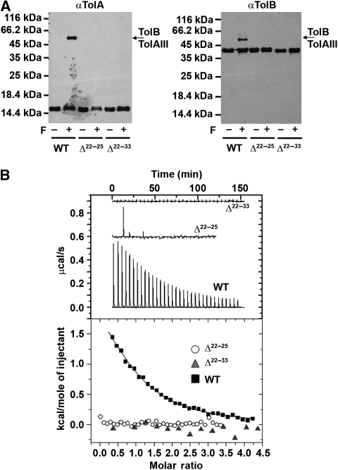
). The cell concentration of the starting complex was 60 μM into which was injected TolAIII. No TolAIII binding could be detected when Pal was bound to TolB. (B) Raw ITC data, top panel and integrated heats of binding for the TolB–TolAIII complex (▪) and the corresponding data when TolB was complexed with the ColE9 T-domain (formula image). Inserts, close-up views of the integrated heats for TolB–Pal + TolAIII, A and for TolB–ColE9 T-domain+TolAIII, (B). See legend to Figure 3 for conditions. Binding curves were fitted to a one-site-binding model using the Origin software. Thermodynamic data are listed in Table II.
Figure 7
Figure 7
TolAIII binding causes the same changes to wild-type TolB 1H-15N TROSY-HSQC spectrum as the deletion of the TolA box. See Materials and methods for details. (A) Overlay of wild-type 15N-TolB (black) and 15N-TolB Δ22−33 (red) spectra. (B) Overlay of wild-type 15N-TolB (black) and 15N-TolB–TolAIII complex (blue) spectra. Circled and boxed regions highlight similarities between the two sets of data. (C) Close-up views of the boxed regions in the two datasets. * indicates equivalent new/shifted peaks in the spectra of TolB as a result of TolAIII binding and deletion of the TolA box.
Figure 8
Figure 8
ColE9 induces disorder in the TolA box. Assignment of TolA box residues for the 75-kDa TolB–ColE9 T-domain complex based on 2D 1H-15N TROSY-HSQC, 3D CBCA(CO)NH and HNCA spectra. See Materials and methods and Supplementary Figure S3 for further details.
Figure 9
Figure 9
Structural model depicting recruitment of TolA by TolB and the subversion of this signalling mechanism by nuclease colicins. (A) In the unbound state, the TolA box of TolB is in conformational equilibrium, most likely alternating between the conformation seen in the Pal-bound state and an intrinsically disordered polypeptide. (B) Binding of the OM lipoprotein Pal (red) to TolB (grey) favours the state in which the TolA box (green) is sequestered to the TolB surface, thereby diminishing association with domain III of TolA, which we assign as the OFF state for the Tol system. (C) In its disordered state, the TolA box recruits TolA, which we assign as the ON state for the Tol system as this couples TolB to TolA and hence to the pmf-linked IM complex of TolQRA. (D) Nuclease colicins such as ColE3–Im3 (shown in the figure; pdb, 1jch) and ColE9–Im9 (this study) use their intrinsically unstructured T-domain TBE to subvert the Tol system by mimicking Pal. Instead of generating the TolB OFF state, however, they promote the disordered ON state of the TolA box. The panel is a composite of several published crystal structures. The colicin translocon comprises the toxin bound to its OM receptor BtuB (pdb, 1ujw), the structure of the ColE3 R-domain bound to BtuB is depicted in the figure (Kurisu et al, 2003), and a porin such as OmpF (pdb, 2zfg) and the periplasmic protein TolB (pdb 1c5k), both of which are recruited by the colicin's intrinsically disordered T-domain (pdb codes, 2zld and 2ivz, respectively). Recruitment of TolAIII (pdb, 1lr0) by the TolA box of TolB generates a ‘trigger complex' that is required for the dissociation of the tightly bound immunity protein of the colicin at the cell surface and initiates translocation across the bacterial OM.

References

    1. Abergel C, Bouveret E, Claverie JM, Brown K, Rigal A, Lazdunski C, Benedetti H (1999) Structure of the Escherichia coli TolB protein determined by MAD methods at 1.95 A resolution. Structure 7: 1291–1300 - PubMed
    1. Baboolal TG, Conroy MJ, Gill K, Ridley H, Visudtiphole V, Bullough PA, Lakey Jeremy H (2008) Colicin N binds to the periphery of its receptor and translocator, outer membrane protein F. Structure 16: 371–379 - PMC - PubMed
    1. Bernadac A, Gavioli M, Lazzaroni JC, Raina S, Lloubes R (1998) Escherichia coli tol-pal mutants form outer membrane vesicles. J Bacteriol 180: 4872–4878 - PMC - PubMed
    1. Bhatt S, Weingart C (2008) Identification of sodium chloride-regulated genes in Burkholderia cenocepacia. Curr Microbiol 56: 418–422 - PubMed
    1. Bonsor DA, Grishkovskaya I, Dodson EJ, Kleanthous C (2007) Molecular mimicry enables competitive recruitment by a natively disordered protein. J Am Chem Soc 15: 4800–4807 - PubMed

Publication types