. 2009;4(9):1295-304.
doi: 10.1038/nprot.2009.127. Epub 2009 Aug 20.

Differentiation of spinal motor neurons from pluripotent human stem cells

Affiliations

Differentiation of spinal motor neurons from pluripotent human stem cells

Bao-Yang Hu et al. Nat Protoc. 2009.

Abstract

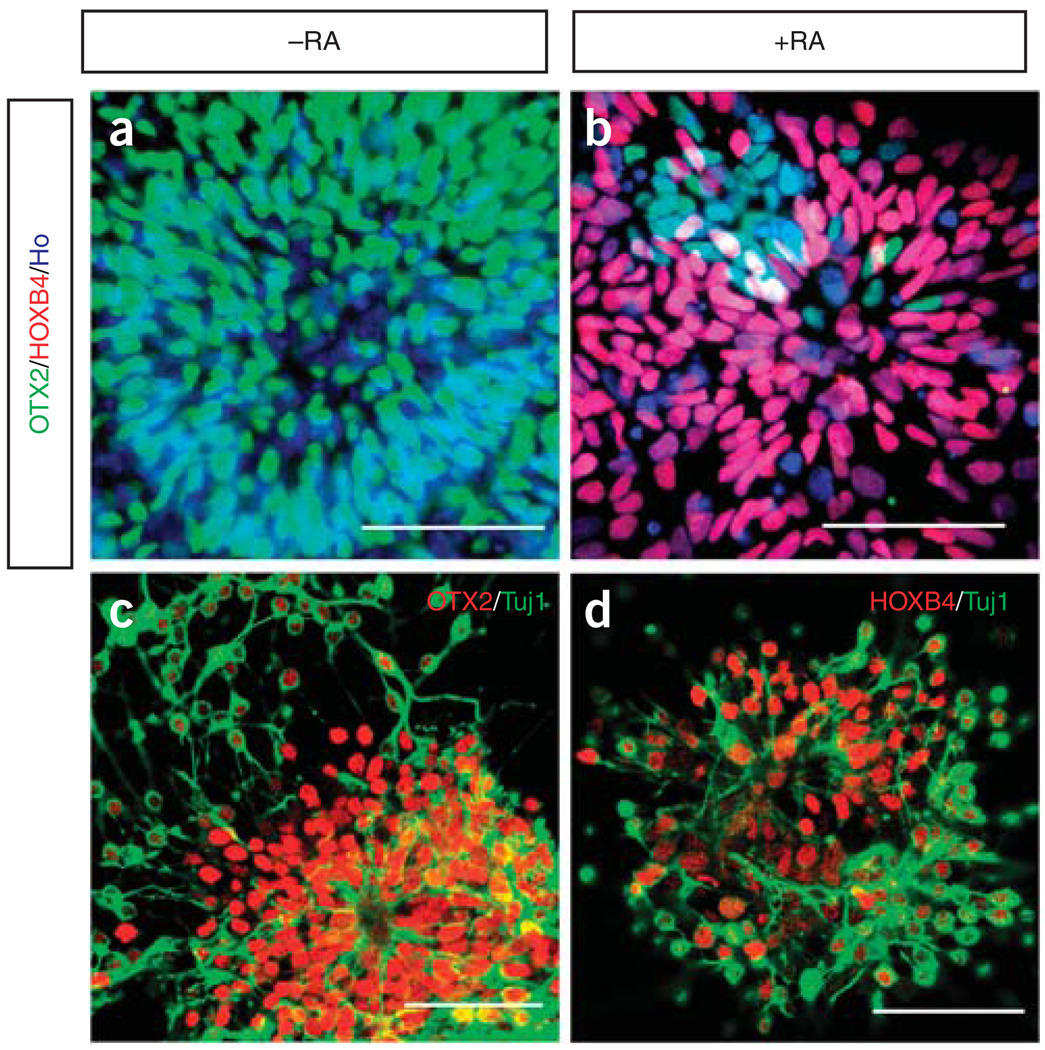
We have devised a reproducible protocol by which human embryonic stem cells (hESCs) or inducible pluripotent stem cells (iPSCs) are efficiently differentiated to functional spinal motor neurons. This protocol comprises four major steps. Pluripotent stem cells are induced to form neuroepithelial (NE) cells that form neural tube-like rosettes in the absence of morphogens in the first 2 weeks. The NE cells are then specified to OLIG2-expressing motoneuron progenitors in the presence of retinoic acid (RA) and sonic hedgehog (SHH) or purmorphamine in the next 2 weeks. These progenitor cells further generate post-mitotic, HB9-expressing motoneurons at the 5th week and mature to functional motor neurons thereafter. It typically takes 5 weeks to generate the post-mitotic motoneurons and 8-10 weeks for the production of functional mature motoneurons. In comparison with other methods, our protocol does not use feeder cells, has a minimum dependence on proteins (purmorphamine replacing SHH), has controllable adherent selection and is adaptable for scalable suspension culture.


Figures

Figure 1
Figure 1
Scheme of differentiation of spinal cord motoneurons (MNs) from hESCs. The starting hESCs are positive for OCT4. After lifting from MEF feeder cells, the hESCs form aggregates (resemble to embryoid bodies, EBs) in suspension. They are cultured for 4 d in hESC medium and in neural differentiation medium thereafter. In the 2nd week, the aggregates that are attached to the surface of culture dishes and possess features of neuroepithelial (NE) rosettes can be enriched for by manual selection. The primitive NE cells (day 10, PAX6-positive) are patterned with retinoic acid (RA) for 5 d. The cells are then PAX6/SOX1-positive and are lifted off the culture surface on day 15 and then cultured with RA and sonic hedgehog (SHH) (or purmorphamine) for the subsequent 2 weeks. The OLIG2-expressing motoneuron progenitors are present in the 4th week. From day 28, these motoneuron progenitors can be differentiated to post-mitotic motoneurons on the substrate in the presence of neurotrophic factors and reduced concentrations of SHH and RA. HB-9-positive neurons form within another week of culture. Except during the initial transition period using the hESC growth medium, the entire process uses a simple serum-free neural differentiation medium. The adherent culture during the neural induction phase is uniquely designed for direct visualization of NE differentiation and for potential purification of the NE cells.
Figure 2
Figure 2
Induction of neuroepithelial cells. hESCs growing on MEF feeders form uniform colonies (a); all cells are positive for OCT4 (red) and negative for PAX6 (green) (b). Columnar epithelial cells at day 10 organize into rosettes in the colony (c); these cells express PAX6 (green) but not SOX1 (red) (d). Neural tube-like rosettes at day 15 (e) stain positive for both PAX6 (green) and SOX1 (red) (f). Bar = 50 µm, Ho shown as blue denotes Hoschest33258-stained nuclei.
Figure 3
Figure 3
Patterning of spinal cord progenitors. The hESC-derived neuroepithelial cells cultured without RA are positive for OTX2 (green) (a). When the cells are cultured in the presence of RA (100 nM) for 1 week, the majority of the cells express HOXB4 (red) (b). (c,d) Following a further week of culture, these progenitors retain the rostral (c) or caudal identity (d) when they differentiate to tubulin+ (Tuj1) (green) neurons. Both OTX2 and HOXB4 are represent as red in c and d. Bar µ 50 µm.
Figure 4
Figure 4
Differentiation and maturation of hESC-derived motoneurons. (a) The motoneuron progenitors are cultured in suspension before they are attached for differentiation. (b) Extensive axonal projections project from the clusters a week after adherent culture. At the 4th week of culture, a large proportion of the cells express OLIG2 (red), whereas a smaller population of HB9-expressing (green) motoneurons is present (c). HB9-positive motorneurons (red) are also positively stained for neuronal marker Tuj1+ (green) at the 5th week (d). At the 6th week of culture, some cells are positive for ChAT (red), a few of the cells co-label with HB9 (green) (e). The hESC-derived motoneurons express synapsin (green) and contact with myotubes in culture (f). The contacts induce clustering of the acetylcholine receptor on myotubes, as shown by bungarotoxin (BTX) (red) staining. Bar = 50 µm in a–e, 20 µm in f.

References

    1. Zhang SC, Wernig M, Duncan ID, Brustle O, Thomson JA. In vitro differentiation of transplantable neural precursors from human embryonic stem cells. Nat. Biotechnol. 2001;19:1129–1133. - PubMed
    1. Li XJ, et al. Specification of motoneurons from human embryonic stem cells. Nat. Biotechnol. 2005;23:215–221. - PubMed
    1. Singh Roy N, et al. Enhancer-specified GFP-based FACS purification of human spinal motor neurons from embryonic stem cells. Exp. Neurol. 2005;196:224–234. - PubMed
    1. Perrier AL, et al. Derivation of midbrain dopamine neurons from human embryonic stem cells. Proc. Natl. Acad. Sci. USA. 2004;101:12543–12548. - PMC - PubMed
    1. Yan Y, et al. Directed differentiation of dopaminergic neuronal subtypes from human embryonic stem cells. Stem Cells. 2005;23:781–790. - PMC - PubMed

Publication types