. 2009 Aug;5(8):e1000553.
doi: 10.1371/journal.ppat.1000553. Epub 2009 Aug 21.

The QseC adrenergic signaling cascade in Enterohemorrhagic E. coli (EHEC)

Affiliations

The QseC adrenergic signaling cascade in Enterohemorrhagic E. coli (EHEC)

David T Hughes et al. PLoS Pathog. 2009 Aug.

Abstract

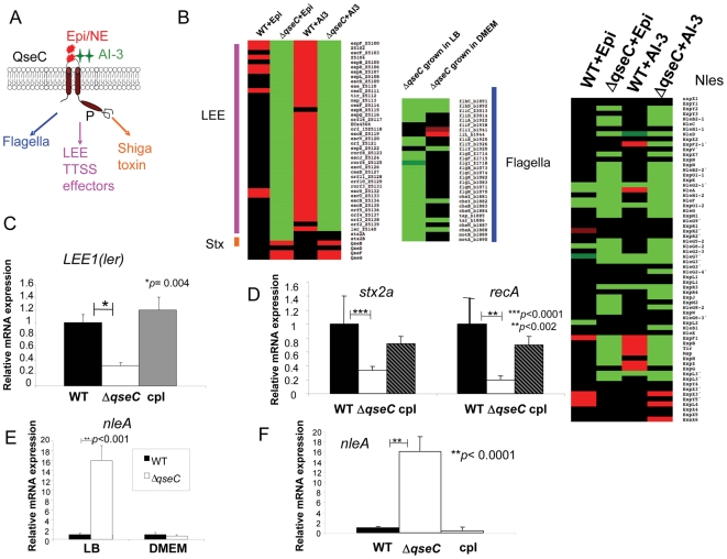
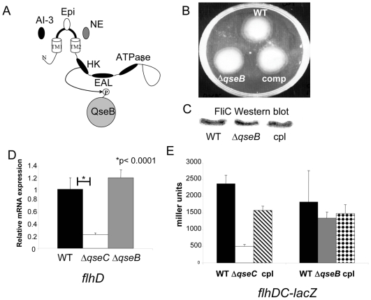
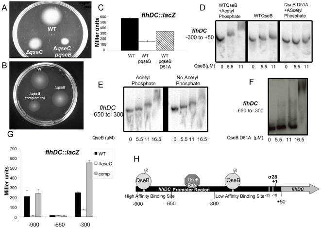
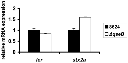
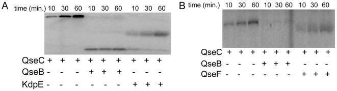
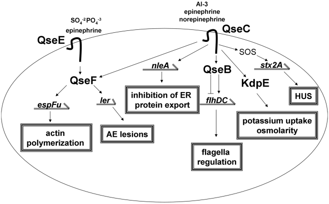
The ability to respond to stress is at the core of an organism's survival. The hormones epinephrine and norepinephrine play a central role in stress responses in mammals, which require the synchronized interaction of the whole neuroendocrine system. Mammalian adrenergic receptors are G-coupled protein receptors (GPCRs); bacteria, however, sense these hormones through histidine sensor kinases (HKs). HKs autophosphorylate in response to signals and transfer this phosphate to response regulators (RRs). Two bacterial adrenergic receptors have been identified in EHEC, QseC and QseE, with QseE being downstream of QseC in this signaling cascade. Here we mapped the QseC signaling cascade in the deadly pathogen enterohemorrhagic E. coli (EHEC), which exploits this signaling system to promote disease. Through QseC, EHEC activates expression of metabolic, virulence and stress response genes, synchronizing the cell response to these stress hormones. Coordination of these responses is achieved by QseC phosphorylating three of the thirty-two EHEC RRs. The QseB RR, which is QseC's cognate RR, activates the flagella regulon which controls bacteria motility and chemotaxis. The QseF RR, which is also phosphorylated by the QseE adrenergic sensor, coordinates expression of virulence genes involved in formation of lesions in the intestinal epithelia by EHEC, and the bacterial SOS stress response. The third RR, KdpE, controls potassium uptake, osmolarity, and also the formation of lesions in the intestine. Adrenergic regulation of bacterial gene expression shares several parallels with mammalian adrenergic signaling having profound effects in the whole organism. Understanding adrenergic regulation of a bacterial cell is a powerful approach for studying the underlying mechanisms of stress and cellular survival.


Conflict of interest statement

The authors have declared that no competing interests exist.

Figures

Figure 1
Figure 1. QseC regulates multiple virulence factors.
(A) Schematic representation of QseC responding to epinephrine/norepinephrine and AI-3 and regulating multiple virulence factors (B) Heat maps from microarray analysis representing the effects of epinephrine and AI-3 on WT EHEC and ΔqseC, differential regulation of the LEE genes, the flagellar genes and the non-LEE encoded secreted effectors are shown. Both WT and ΔqseC produce AI-3 in DMEM (OD600 1.0), hence these data reflect the transcriptome in the presence of AI-3 alone (WT+AI-3 and ΔqseC+AI-3) or AI-3 plus epinephrine (WT+Epi, ΔqseC+Epi) (C) QPCR of ler in wt EHEC, ΔqseC, and ΔqseC complement strain grown in DMEM (OD600 1.0) (in the presence of self produced AI-3) (D) QPCR of stx2a and recA in wt EHEC, ΔqseC, and ΔqseC complement strain grown in DMEM (OD600 1.0) (in the presence of self produced AI-3) (E) QPCR of nleA in WT EHEC and ΔqseC in LB and DMEM (OD600 1.0) (in the presence of self produced AI-3) (F) QPCR of nleA in wt EHEC, ΔqseC, and ΔqseC complement strain grown in DMEM (OD600 1.0) (in the presence of self produced AI-3).
Figure 2
Figure 2. ΔqseC and ΔqseB do not have the same phenotype.
(A) Schematic representation of QseC responding to the signals epinephrine/norepinephrine and AI-3 and transferring its phosphate onto its cognate response regulator QseB. (B) Motility plate of wt EHEC, ΔqseB, and the ΔqseB complement strain (complemented with plasmid pVS178, qseBC in pBAD33 [35]) (in the presence of self produced AI-3) (C) Western blot of FliC in wt EHEC, ΔqseB, and the ΔqseB complement strain (complemented with plasmid pVS178, qseBC in pBAD33) (in the presence of self produced AI-3) (D) QPCR of flhD in wt EHEC, ΔqseC, and ΔqseB in LB (OD600 1.0) (in the presence of self produced AI-3) (E) β-galactosidase assay of the flhDC promoter controlling lacZ expression in wt EHEC, ΔqseC, the ΔqseC complement strain, ΔqseB, and the ΔqseB complement strain (complemented with plasmid pVS178, qseBC in pBAD33) in LB (OD600 1.0) (in the presence of self produced AI-3).
Figure 3
Figure 3. QseB differentially regulates flhDC based on its phosphorylation state.
(A) Motility plate of wt EHEC, ΔqseC, and ΔqseC overexpressing qseB (in the presence of self produced AI-3) (B) Motility plate of wt EHEC, ΔqseB, and ΔqseB overexpressing qseB (in the presence of self produced AI-3) (C) β-galactosidase assay of the flhDC promoter controlling lacZ expression in wt EHEC and in wt EHEC overexpressing qseB and the QseB D51A mutant in LB (OD600 1.0) (in the presence of self produced AI-3) (D) EMSA of the −300 bp to +50 bp region of the flhDC promoter with QseB and the phosphor-donor acetyl phosphate, QseB, and the nonphosphorylatable QseB D51A with the phosphor-donor acetyl phosphate (E) EMSA of the −50 bp to +50 bp region of the flhDC promoter with QseB in the presence and absence of the phosphor-donor acetyl phosphate (F) EMSA of the −650 bp to +50 bp region of the flhDC promoter with the nonphosphorylatable QseB D51A (G) Nested deletion β-galactosidase analysis of the flhDC promoter (−900 bp to +50 bp, −650 bp to +50 bp, and −300 bp to +50 bp) controlling lacZ expression in wt EHEC, ΔqseC, the ΔqseC complement strain in LB (OD600 1.0) (in the presence of self produced AI-3) (H) QseB binding sites on the flhDC promoter.
Figure 4
Figure 4. qPCR of ler and stx2a in wt EHEC and ΔqseB in DMEM (OD600 1.0) (in the presence of self produced AI-3).
Figure 5
Figure 5. QseC phosphotransfers to the response regulators QseB, QseF and KdpE.
(A) Autoradiograph of QseC autophosphorylation in lipid vesicles in the presence of 10 µM epinephrine and phosphotransfer onto QseB and KdpE (B) Autoradiograph of QseC autophosphorylation in lipid vesicles in the presence of 10 µM epinephrine and phosphotransfer onto QseB and QseF.
Figure 6
Figure 6. QseC, KdpE and QseF regulatory targets.
(A) QPCR of kdpA in wt EHEC, ΔqseC, and ΔqseC complement strain in DMEM (OD600 1.0) (in the presence of self produced AI-3) (B) QPCR of flhD in wt EHEC, ΔqseC, and ΔkdpE in DMEM (OD600 1.0) (in the presence of self produced AI-3) (C) Motility plate of wt EHEC, ΔqseB, ΔkdpE, and ΔqseC (D) QPCR of ler and stx2a in wt EHEC and ΔkdpE in DMEM (OD600 1.0) (in the presence of self produced AI-3) (E) QPCR of ler and stx2a in wt EHEC and ΔqseF in DMEM (OD600 1.0) (in the presence of self produced AI-3).
Figure 7
Figure 7. Regulatory overlap of QseC and its phosphorylation targets.
(A) Schematic representation of QseC responding to the signals epinephrine/norepinephrine and AI-3 and transferring its phosphate onto QseB, KdpE, and QseF (B) Microarray analysis comparing ΔqseC to ΔqseB, ΔkdpE, and ΔqseF in DMEM (OD600 1.0) (in the presence of self produced AI-3).
Figure 8
Figure 8. Model of the QseC and QseE signaling cascades in EHEC.

References

    1. Molina PE. Endocrine Physiology: The McGraw Hill Companies Inc; 2006.
    1. Sperandio V, Torres AG, Jarvis B, Nataro JP, Kaper JB. Bacteria-host communication: the language of hormones. Proc Natl Acad Sci USA. 2003;100:8951–8956. - PMC - PubMed
    1. Lyte M, Ernst S. Catecholamine induced growth of gram negative bacteria. Life Sci. 1992;50:203–212. - PubMed
    1. Clarke MB, Hughes DT, Zhu C, Boedeker EC, Sperandio V. The QseC sensor kinase: a bacterial adrenergic receptor. Proc Natl Acad Sci U S A. 2006;103:10420–10425. - PMC - PubMed
    1. Reading NC, Rasko DA, Torres AG, Sperandio V. The two-component system QseEF and the membrane protein QseG link adrenergic and stress sensing to bacterial pathogenesis. Proc Natl Acad Sci U S A. 2009;106:5889–5894. - PMC - PubMed

Publication types

MeSH terms