. 2009 Sep 15;81(18):7823-8.
doi: 10.1021/ac901176t.

Preferentially increased nitration of alpha-synuclein at tyrosine-39 in a cellular oxidative model of Parkinson's disease

Affiliations

Preferentially increased nitration of alpha-synuclein at tyrosine-39 in a cellular oxidative model of Parkinson's disease

Steven R Danielson et al. Anal Chem. .

Abstract

Alpha-synuclein is a major component of Lewy bodies, proteinacious inclusions which are a major hallmark of Parkinson's disease (PD). Lewy bodies contain high levels of nitrated tyrosine residues as determined by antibodies specific for 3-nitrotyrosine (3NT) and via mass spectrometry (MS). We have developed a multiple reaction monitoring (MRM) mass spectrometry method to sensitively quantitate the 3NT levels of specific alpha-synuclein tyrosine residues. We found a 9-fold increase (relative to controls) in levels of 3NT at Tyr-39 of alpha-synuclein in an inducible transgenic cellular model of Parkinson's disease in which monoamine oxidase B (MAO-B) is overexpressed and which emulates several features of PD. Increased nitration of Tyr-39 on endogenous alpha-synuclein via elevations in MAO-B levels could be abrogated by the addition of deprenyl, a specific MAO-B inhibitor. The increased levels of 3NT was selective for Tyr-39 as no significant increases in 3NT levels were detected at other tyrosine residues present in the protein (Tyr-125, Tyr-133, and Tyr-136). This is the first report of increased 3NT levels of a specific tyrosine in a PD model and the first use of MRM mass spectrometry to quantify changes in 3NT modifications at specific sites within a target protein.


Figures
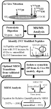
Figure 1
Figure 1
Diagram depicting summarized strategy to identify and quantify PTMs (3NT) on low abundant peptides, starting with in vitro nitrated (tetranitromethane) recombinant α-synuclein which was subsequently digested and analyzed by MS/MS to identify 3NT modified peptides using rigorous criteria described in the Results section. Optimized MRM transitions were then used to quantify 3NT modified and unmodified peptides present on endogenous α-synuclein immunoprecipitated (IP) from a cellular model of PD using the equation listed in the final step titled MRM analysis (TIC = Total Ion Chromatogram).
Figure 2
Figure 2
Identification of α-synuclein peptide E-35-GVLYNO2VGSKTK-45 containing 3NT modification at Tyr-39. Human α-synuclein was in vitro nitrated, proteolytically digested with Asp-N and then analyzed by MS/MS. A) MS/MS spectra of unmodified human α-synuclein peptide (Asp-N digested) containing Tyr-39, E-35-GVLYVGSKTK-45 at m/z 394.2, (M=1179.6 Da). B) Corresponding MS/MS spectra of the peptide containing 3NT, E-35-GVLYNO2VGSKTK-45 (YNO2 = 3NT) at m/z 409.2 (M=1224.5 Da). Note the low mass immonium (IMM) ions (+NH2=CH-R), for Tyr and 3NT at m/z 136.1 and 181.1, respectively.
Figure 3
Figure 3
Chromatographic retention time shift of 3NT modified peptide. MRM total ion chromatogram of unmodified and 3NT modified peptide (Asp-N digest) containing Tyr-39 showing retention time delay of ~2 min for the nitrated peptide relative to the unmodified form. Four MRM transitions were used each for the unmodified and 3NT modified peptides, all of which are listed in Table S-2 (Supporting Information).
Figure 4
Figure 4
Increased 3NT levels at Tyr-39 of endogenous α-synuclein due to MAO-B overexpression in dopaminergic PC12 cells as determined by MRM (4000 QTRAP). A 9-fold increase of 3NT levels at Tyr-39 was observed in PC12 cells overexpressing MAO-B relative to control levels. This increase was abrogated by the addition of the MAO-B inhibitor deprenyl. As a positive control, cells were treated with 150 μM of H2O2 for 24 hours which also exhibited a large increase (6-fold) in 3NT levels as compared to control cells. Quantitation was done using the MRM transitions (Q1/Q3): unmodified 394.2/520.3 and 3NT modified 409.2/520.3. Error bars are the standard deviation of 3 biological replicates analyzed in singlicate using MRM transitions for unmodified and 3NT modified peptides.

References

    1. Polymeropoulos MH, Lavedan C, Leroy E, Ide SE, Dehejia A, Dutra A, Pike B, Root H, Rubenstein J, Boyer R, Stenroos ES, Chandrasekharappa S, Athanassiadou A, Papapetropoulos T, Johnson WG, Lazzarini AM, Duvoisin RC, Di Iorio G, Golbe LI, Nussbaum RL. Science. 1997;276:2045–2047. - PubMed
    1. Baba M, Nakajo S, Tu PH, Tomita T, Nakaya K, Lee VM, Trojanowski JQ, Iwatsubo T. Am J Pathol. 1998;152:879–884. - PMC - PubMed
    1. Good PF, Hsu A, Werner P, Perl DP, Olanow CW. J Neuropathol Exp Neurol. 1998;57:338–342. - PubMed
    1. Giasson BI, Duda JE, Murray IV, Chen Q, Souza JM, Hurtig HI, Ischiropoulos H, Trojanowski JQ, Lee VM. Science. 2000;290:985–989. - PubMed
    1. Przedborski S, Chen Q, Vila M, Giasson BI, Djaldatti R, Vukosavic S, Souza JM, Jackson-Lewis V, Lee VM, Ischiropoulos H. J Neurochem. 2001;76:637–640. - PubMed

Publication types