Review
. 2010 Apr;16(2):169-87.
doi: 10.1089/ten.TEB.2009.0352.

Challenges in cardiac tissue engineering

Affiliations
Review

Challenges in cardiac tissue engineering

Gordana Vunjak-Novakovic et al. Tissue Eng Part B Rev. 2010 Apr.

Abstract

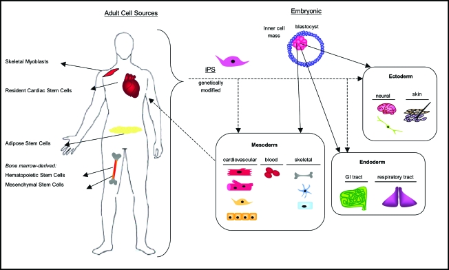
Cardiac tissue engineering aims to create functional tissue constructs that can reestablish the structure and function of injured myocardium. Engineered constructs can also serve as high-fidelity models for studies of cardiac development and disease. In a general case, the biological potential of the cell-the actual "tissue engineer"-is mobilized by providing highly controllable three-dimensional environments that can mediate cell differentiation and functional assembly. For cardiac regeneration, some of the key requirements that need to be met are the selection of a human cell source, establishment of cardiac tissue matrix, electromechanical cell coupling, robust and stable contractile function, and functional vascularization. We review here the potential and challenges of cardiac tissue engineering for developing therapies that could prevent or reverse heart failure.


Figures

FIG. 1.
FIG. 1.
Cell sources for cardiac regeneration. A variety of both adult and embryonic sources have emerged as candidates for cell therapy. Skeletal myoblasts, the first to be seen in the clinical setting, have been followed by bone marrow–derived mesenchymal and hematopoetic stem cells, and recently adipose-derived stem cells. Of these nonmyogenic somatic cells, only resident cardiac stem cells have been identified as endogenous myogenic progenitors, but their limited number eliminates them as a reliable candidate in cell therapy. Embryonic stem cells, derived from the inner cell mass of the blastocyst, give rise to all the three germ layers (endoderm, mesoderm, ectoderm) and their various cell populations. Successful derivation of cardiovascular lineages from embryonic stem cells has presented them as the most attractive myogenic source for cell therapy. Recent success with induced pluripotent stem (iPS) cells, genetically modified adult cells that display “embryonic-like” pluripotency, have hinted at their potential to differentiate into cardiovascular lineages. iPS cells represent a promising myogenic source of cardiac cells for heart therapy, eluding both ethical and immunogenic concerns, but raising questions regarding chromosomal stability. Color images available online at www.liebertonline.com/ten.
FIG. 2.
FIG. 2.
Delivery to the heart—cells or tissues? Existing administration of liquid-phase cell suspensions suffers from low engraftment and cell survival. Alternative solutions for cell delivery are investigated to provide control over cell retention in the injury site, survival, and coupling with the host cells. Hydrogel cell delivery via catheters may aid to cell retention and also provide environmental conditioning via hydrogel design. Tissue engineering of a contractile cardiac patch seeks to deliver cells within a tissue-like microenvironment with immediate function and a capacity for vascular and electromechanical integration with the heart. Color images available online at www.liebertonline.com/ten.
FIG. 3.
FIG. 3.
The basic methods of three-dimensional (3D) stem-cell culture (cell seeding onto 3D scaffolds and bioreactor culture) are well established, but the challenge of studying stem cells under conditions predictive of their behavior in vivo remains. By carefully studying various characteristics of the native (developing or adult) cell environment and applying them during 3D culture (with scaffolds and/or bioreactors), one can begin to develop differentiated, functional tissues from human stem-cell sources. Color images available online at www.liebertonline.com/ten.
FIG. 4.
FIG. 4.
Current concepts in cardiac tissue engineering. Several approaches have been used to generate 3D cardiac patches in vitro, including the creation of stackable cell sheets without using a scaffold, the use of molds to create constructs of cells cultured in hydrogel, and the culture of cells cultured on porous scaffolds. These constructs may then be subjected to physical stimuli (such as electrical excitation, mechanical stretch, and medium flow) via scaffolds and bioreactors. The major hurdle continues to be finding ways to provide vascularization/blood supply to the cardiac cells. Likely, an approach delivering a combination of relevant factors will need to be used to yield a functional, implantable construct. Color images available online at www.liebertonline.com/ten.

References

    1. Harrison R.G. Observations on the living developing nerve fiber. Proc Soc Exp Biol Med. 1907;4:140.
    1. Langer R. Vacanti J.P. Tissue Eng Sci. 1993;260:920. - PubMed
    1. Lanza R. Langer R. Vacanti J. Principles of Tissue Engineering. Boston: Elsevier Academic Press; 2007.
    1. Severs N.J. The cardiac muscle cell. Bioessays. 2000;22:188. - PubMed
    1. American Heart Association Cardiovascular Disease Statistics. 2008.

Publication types

LinkOut - more resources