Review
. 2009 Aug 27;422(3):393-403.
doi: 10.1042/BJ20090978.

Molecular biology, genetics and biochemistry of the repulsive guidance molecule family

Affiliations
Review

Molecular biology, genetics and biochemistry of the repulsive guidance molecule family

Christopher J Severyn et al. Biochem J. .

Abstract

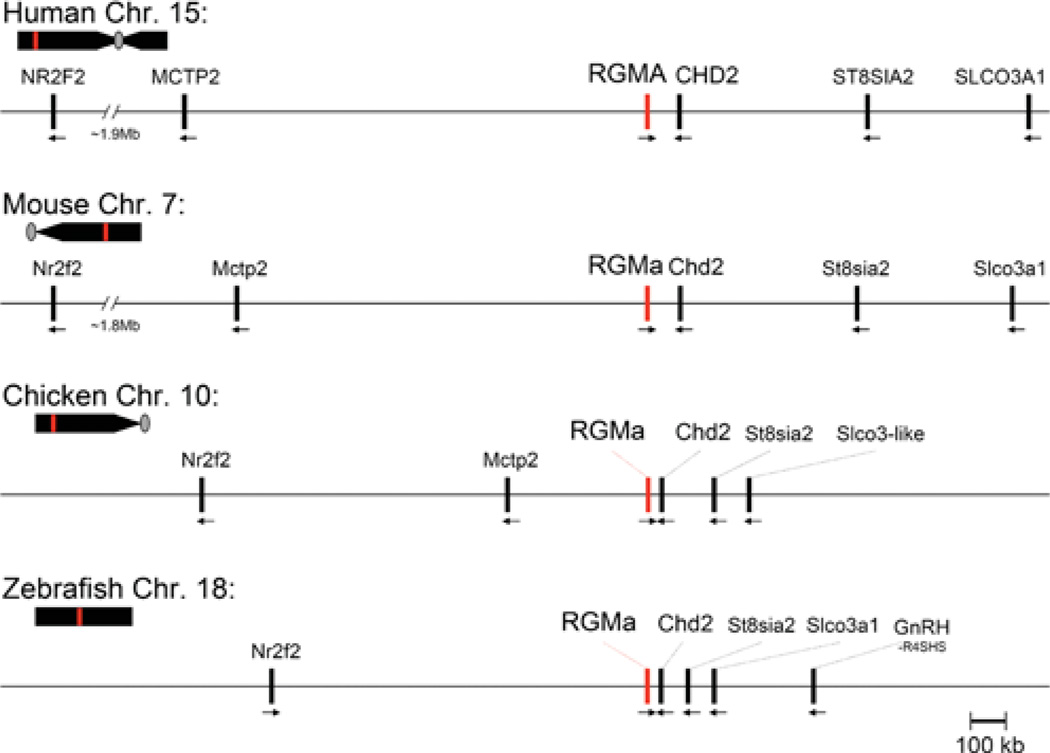
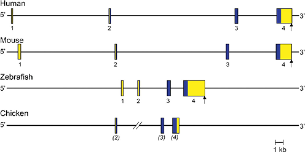
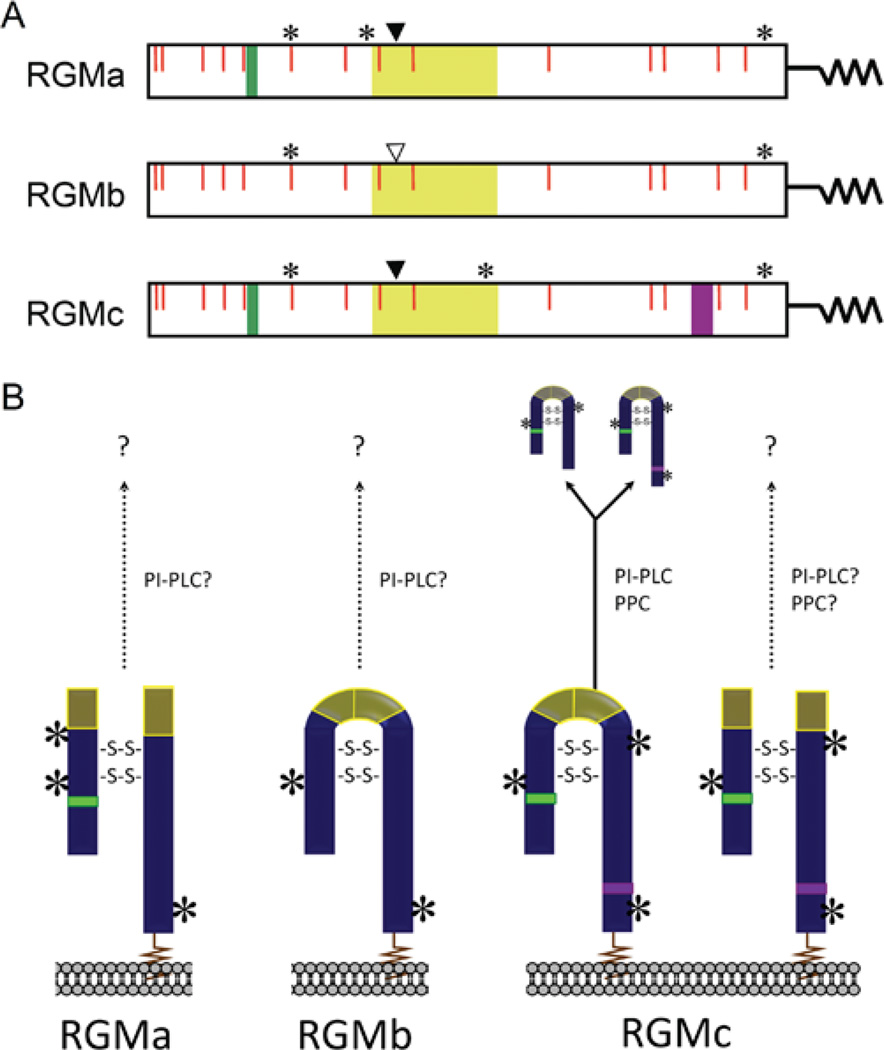
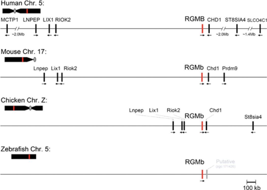
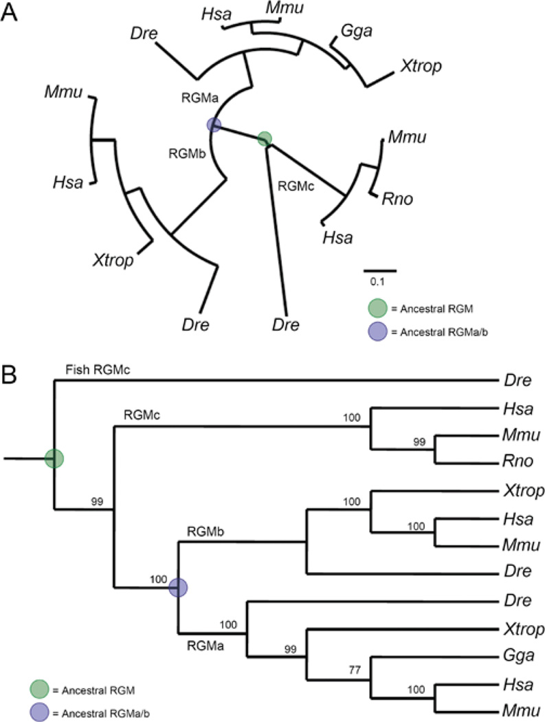
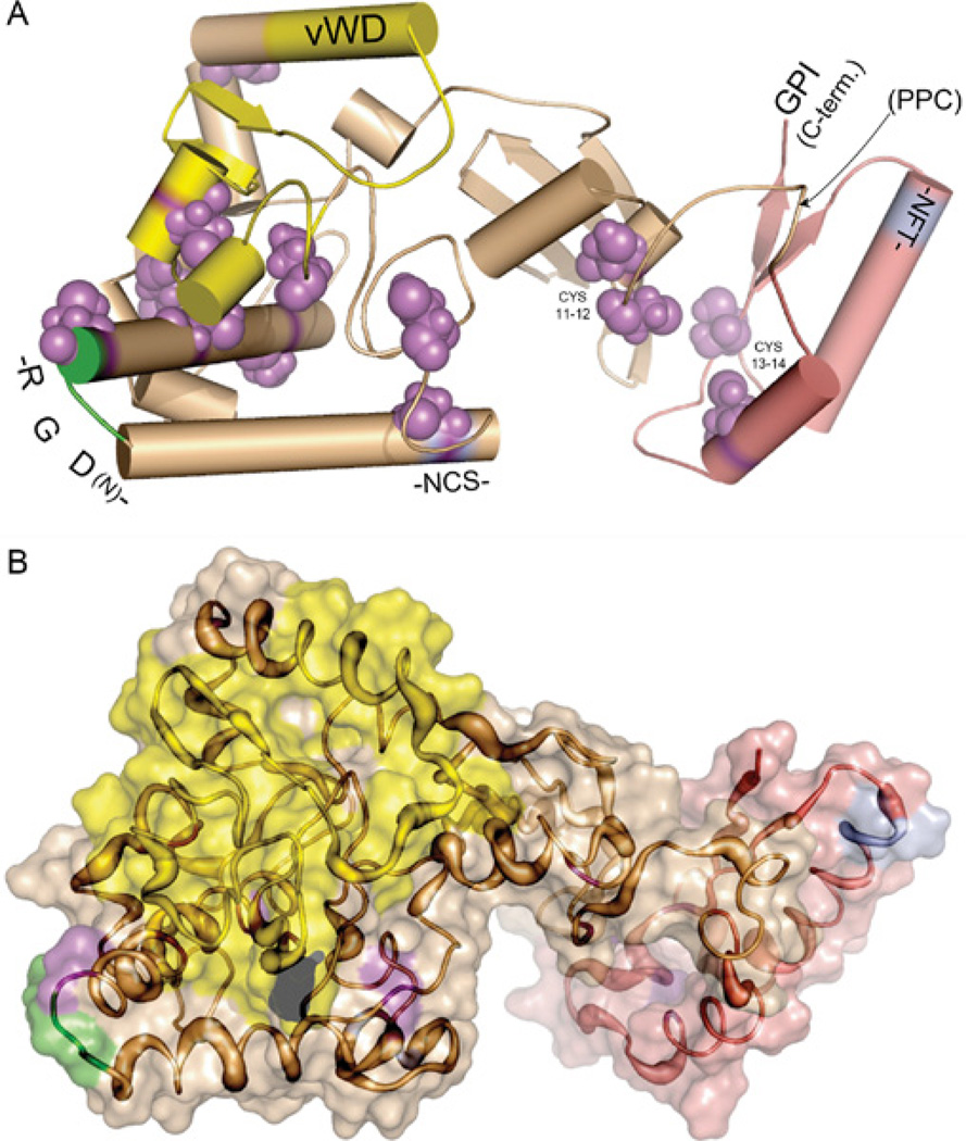
RGMs (repulsive guidance molecules) comprise a recently discovered family of GPI (glycosylphosphatidylinositol)-linked cell-membrane-associated proteins found in most vertebrate species. The three proteins, RGMa, RGMb and RGMc, products of distinct single-copy genes that arose early in vertebrate evolution, are approximately 40-50% identical to each other in primary amino acid sequence, and share similarities in predicted protein domains and overall structure, as inferred by ab initio molecular modelling; yet the respective proteins appear to undergo distinct biosynthetic and processing steps, whose regulation has not been characterized to date. Each RGM also displays a discrete tissue-specific pattern of gene and protein expression, and each is proposed to have unique biological functions, ranging from axonal guidance during development (RGMa) to regulation of systemic iron metabolism (RGMc). All three RGM proteins appear capable of binding selected BMPs (bone morphogenetic proteins), and interactions with BMPs mediate at least some of the biological effects of RGMc on iron metabolism, but to date no role for BMPs has been defined in the actions of RGMa or RGMb. RGMa and RGMc have been shown to bind to the transmembrane protein neogenin, which acts as a critical receptor to mediate the biological effects of RGMa on repulsive axonal guidance and on neuronal survival, but its role in the actions of RGMc remains to be elucidated. Similarly, the full spectrum of biological functions of the three RGMs has not been completely characterized yet, and will remain an active topic of ongoing investigation.


Figures

Figure 1
Figure 1. Comparative structures of RGMa genomic loci
The relative position of the RGMa gene (red line) is indicated on each chromosome (Chr.; human 15, mouse 7, chicken 10, zebrafish 18) in relation to the centromere (grey oval, if information available) and telomere. Presented below each chromosome is a higher resolution view of the RGMa locus for each species. Neighbouring genes are indicated, with the transcriptional direction represented by an arrow. Gene names corresponding to the abbreviations may be found in Table 4.
Figure 2
Figure 2. Comparative organization of RGMa genes
The anatomy of human, mouse, zebrafish and chicken RGMa genes is shown. Exons are indicated by boxes, with coding regions in blue and non-coding regions in yellow. The assignment of exon numbers is based on comparison with mouse RGMa. The polyadenylation site, when known, is depicted by a vertical arrow. The location of zebrafish exon 1 is based on mapping available EST (expressed sequence tag) data taken from GenBank® (accession numbers AL911518 and EH589480). The length of one of the introns of chicken RGMa is not known (shown as two angled lines), as the putative exon 2 cannot be mapped to the genomic DNA sequence, which appears to be incomplete in this region. Chicken exon assignments are in parentheses because the putative exon 1 cannot be mapped to the genome.
Figure 3
Figure 3. Characteristics of RGM proteins
(A) The linear maps of mature RGMa, RGMb and RGMc contain the following features: RGD motif (RGMa and RGMc, green); vWD, partial vWD domain (yellow); PPC, PPC recognition and cleavage site (RGMc only, purple); *location of asparagine-linked glycosylation sites; solid arrowhead, site of intra-molecular proteolytic cleavage to generate two-chain RGMa and RGMc; vertical open arrowhead, possible site of intra-molecular proteolytic cleavage in RGMb; red vertical lines, conserved cysteine residues. The squiggle at the C-terminus of each protein represents the GPI anchor. (B) Schematic of mature RGMa, RGMb and RGMc on the cell surface, as well as the secreted forms of RGMc. Based on published studies, RGMa appears to be primarily a two-chain molecule, and RGMb a single-chain protein, whereas RGMc appears to be represented by both single- and two-chain species. Experimental data supports at least one disulfide bond between the N- and C-termini [9,45,47], and ab inito molecular modelling (see Figure 9) predicts one or two disulfide bonds connecting the two-chain RGM isoforms (shown as -S-S-), though the exact number is currently unknown. Single chain RGMc is released from the cell surface, and is found in extracellular fluid and in blood [,–48], potentially through the actions of a furin-like PPC and/or a PI-PLC. It is not known if RGMa, RGMb or two-chain RGMc are released from the membrane (as indicated by arrows with question marks). Locations of asparagine-linked glycosylation sites are indicated by asterisks, and the GPI anchor is depicted as a squiggle.
Figure 4
Figure 4. Comparative structures of RGMb genomic loci
The relative position of the RGMb gene (red line) is indicated on each chromosome (Chr.; human 5, mouse 17, chicken Z, zebrafish 5) in relation to the centromere (grey oval, if information available) and telomere. Presented below each chromosome is a higher resolution view of the RGMb locus for each species. Neighbouring genes are indicated, with their transcriptional direction represented by an arrow. For the zebrafish RGMb locus, a nearby provisional gene is shown in grey; to date no other genes have been mapped to this region. Gene names corresponding to the abbreviations may be found in Table 4.
Figure 5
Figure 5. Comparative organization of RGMb genes
The anatomy of human, mouse and zebrafish RGMb genes is shown. The assignment of exon numbers is based on comparison with human RGMb, and is provisional for mouse and zebrafish, as indicated by the parentheses. Exons are indicated by boxes, with coding regions in blue and non-coding regions in yellow. The polyadenylation site, when known, is depicted by a vertical arrow. Only coding information is available for zebrafish RGMb.
Figure 6
Figure 6. Comparative structures of RGMc genomic loci
The relative position of the RGMc gene (red line) is indicated on each chromosome (Chr.; human 1, mouse 3, zebrafish 16) in relation to the centromere (grey oval, if information available) and telomere. Presented below each chromosome is a higher resolution view of the RGMc locus for each species. Neighbouring genes are indicated, with their transcriptional direction represented by an arrow. Lix1-like, shown in grey, is a putative pseudo-gene (Lix1l), as there is no known transcript available in GenBank®. Gene names corresponding to the abbreviations may be found in Table 4.
Figure 7
Figure 7. Comparative organization of RGMc genes
The anatomy of human, mouse and zebrafish RGMc genes is shown. Exons are indicated by boxes, with coding regions in blue and non-coding regions in yellow. The assignment of exon numbers is based on comparison with mouse RGMc, and is provisional for zebrafish (in parentheses). The polyadenylation site is represented by a vertical arrow.
Figure 8
Figure 8. Phylogeny of the RGM family
Evolutionary trees have been derived from the protein translation of well-annotated RGM DNA sequences in which the mRNA and gene agrees. Methods of analysis are as follows: seven separate MSAs (multiple sequence alignments) of full-length RGM proteins were performed with MUSCLE [14], Clustal-W [75] or hand alignment, followed by direct submission or a codon-optimized alignment through PAL2NAL [76]. Either protein MSAs or codon-based alignments were submitted to several phylogenetic methods, including neighbour joining with unrooted and rooted trees (via MacVector), maximum likelihood [77,78] (with and without Bootstrap methods on neighbour joining and maximum likelihood) and Bayesian [79,80] analysis. (A) RGM family phylogeny using an unrooted maximum likelihood method, displaying a distance of 0.1 amino acid substitutions per position (scale bar). (B) RGM family cladogram derived from the neighbour joining method (Poisson-correction with gaps distributed proportionally) rooted with zebrafish (Dre) RGMc, displaying bootstrap values as percentage of 5000 replications supporting that branch on the cladogram. Species abbreviations for (A) and (B) may be found in Table 1. For both (A) and (B), the putative ancestral RGM is highlighted in green and the ancestral gene to RGMa and RGMb is shown in blue. Phylogeny and cladogram created using Pal2NAL [76], Selection Server [81], Phylogeny.fr [78], PhyML 3.0 [77], TreeDyn [82] and MacVector v7.2.3.
Figure 9
Figure 9. Ab initio model for RGM proteins
The model was generated using Rosetta [–67,70,83], using the following steps: First, 1000 independent structures were predicted from a fragment library prepared with the Robetta Fragment server [63,68,69]. Structures were clustered for similarity based on their root mean square deviations. The centres of the three largest clusters were chosen as the best models, defined as having the lowest standard deviation of the mean among positions of carbon atoms of all residues to all other simulations in a cluster. Selected structures were minimized using CharmM [84,85] and analysed for consistency with known experimental data as described in [86]. A single model is illustrated. (A) Cartoon version of the model. Cylinders represent α-helical regions, thick lines with arrows represent β-sheets, and thin lines represent unstructured regions. The model suggests that members of the RGM family adopt a two-lobe structure. The RGD domain is depicted in green, the partial vWD domain is in yellow, cysteines are in purple, asparagine-linked glycosylation sites conserved in all 3 mammalian RGMs are in cyan (and labeled -NCS- and -NFT-), and the GPI anchor attachment site at the C-terminus (C-term.) is noted. All of the above regions appear to be surface exposed. The PPC site (found only in mammalian RGMc) is depicted by a labelled arrow. The N-terminus is not visible as it is located behind the partial vWD domain in the left lobe of the protein. An interactive three-dimensional version of (A) can be found at http://www.BiochemJ.org/422/0393/bj4220393add.him. (B) Space-filling version of the model. The increasing thickness of the tubes represents greater divergence in primary amino acid sequences among RGM family members. The protein domains are colour-coded as in (A).

References

    1. Niederkofler V, Salie R, Sigrist M, Arber S. Repulsive guidance molecule (RGM) gene function is required for neural tube closure but not retinal topography in the mouse visual system. J. Neurosci. 2004;24:808–818. - PMC - PubMed
    1. Monnier PP, Sierra A, Macchi P, Deitinghoff L, Andersen JS, Mann M, Flad M, Hornberger MR, Stahl B, Bonhoeffer F, Mueller BK. RGM is a repulsive guidance molecule for retinal axons. Nature. 2002;419:392–395. - PubMed
    1. Schmidtmer J, Engelkamp D. Isolation and expression pattern of three mouse homologues of chick Rgm. Gene Expr. Patterns. 2004;4:105–110. - PubMed
    1. Samad TA, Srinivasan A, Karchewski LA, Jeong SJ, Campagna JA, Ji RR, Fabrizio DA, Zhang Y, Lin HY, Bell E, Woolf CJ. DRAGON: a member of the repulsive guidance molecule-related family of neuronal- and muscle-expressed membrane proteins is regulated by DRG11 and has neuronal adhesive properties. J. Neurosci. 2004;24:2027–2036. - PMC - PubMed
    1. Kuninger D, Kuzmickas R, Peng B, Pintar JE, Rotwein P. Gene discovery by microarray: identification of novel genes induced during growth factor-mediated muscle cell survival and differentiation. Genomics. 2004;84:876–889. - PubMed

Publication types