. 2009 Aug 22:9:55.
doi: 10.1186/1472-6807-9-55.

The crystal structure of Haloferax volcanii proliferating cell nuclear antigen reveals unique surface charge characteristics due to halophilic adaptation

Affiliations

The crystal structure of Haloferax volcanii proliferating cell nuclear antigen reveals unique surface charge characteristics due to halophilic adaptation

Jody A Winter et al. BMC Struct Biol. .

Abstract

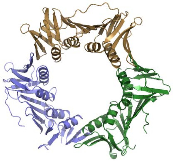
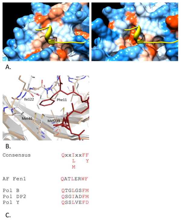
Background: The high intracellular salt concentration required to maintain a halophilic lifestyle poses challenges to haloarchaeal proteins that must stay soluble, stable and functional in this extreme environment. Proliferating cell nuclear antigen (PCNA) is a fundamental protein involved in maintaining genome integrity, with roles in both DNA replication and repair. To investigate the halophilic adaptation of such a key protein we have crystallised and solved the structure of Haloferax volcanii PCNA (HvPCNA) to a resolution of 2.0 A.

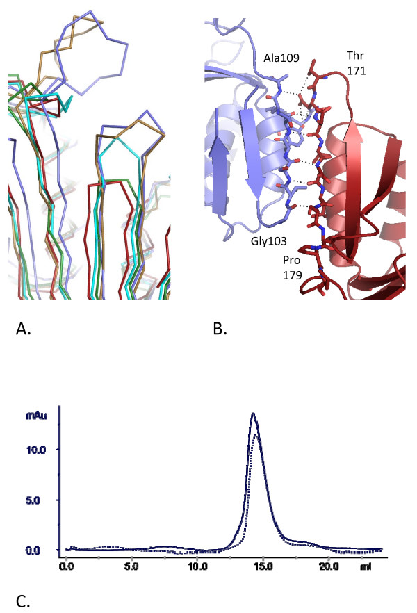
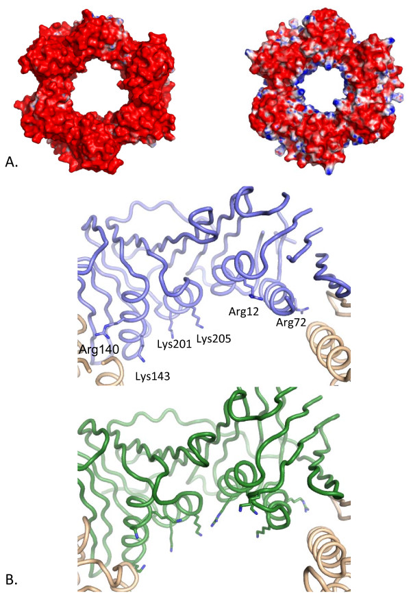
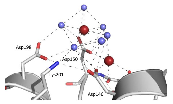
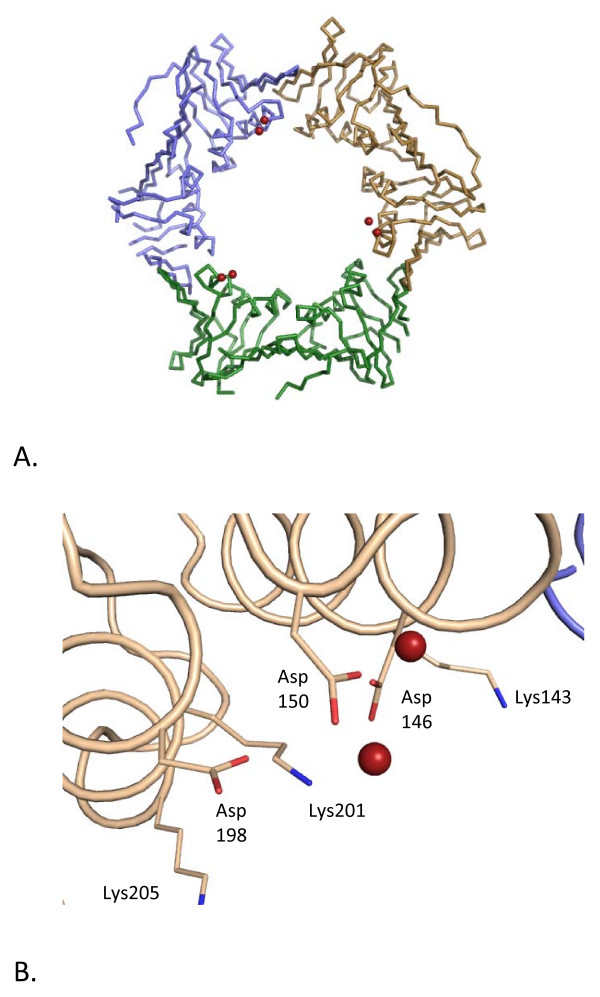
Results: The overall architecture of HvPCNA is very similar to other known PCNAs, which are highly structurally conserved. Three commonly observed adaptations in halophilic proteins are higher surface acidity, bound ions and increased numbers of intermolecular ion pairs (in oligomeric proteins). HvPCNA possesses the former two adaptations but not the latter, despite functioning as a homotrimer. Strikingly, the positive surface charge considered key to PCNA's role as a sliding clamp is dramatically reduced in the halophilic protein. Instead, bound cations within the solvation shell of HvPCNA may permit sliding along negatively charged DNA by reducing electrostatic repulsion effects.

Conclusion: The extent to which individual proteins adapt to halophilic conditions varies, presumably due to their diverse characteristics and roles within the cell. The number of ion pairs observed in the HvPCNA monomer-monomer interface was unexpectedly low. This may reflect the fact that the trimer is intrinsically stable over a wide range of salt concentrations and therefore additional modifications for trimer maintenance in high salt conditions are not required. Halophilic proteins frequently bind anions and cations and in HvPCNA cation binding may compensate for the remarkable reduction in positive charge in the pore region, to facilitate functional interactions with DNA. In this way, HvPCNA may harness its environment as opposed to simply surviving in extreme halophilic conditions.


Figures

Figure 1
Figure 1
Architecture of HvPCNA. Cartoon representation of the HvPCNA trimer. Chain A is shown in purple, chain B in green and chain C in gold.
Figure 2
Figure 2
Interactions at the monomer-monomer interface. A. Superposition of known PCNA structures, showing variation in the extent of the monomer-monomer interface. Red – H. volcanii; green – A. fulgidus [PDB:1RWZ]; cyan – P. furiosus [PDB:1GE8]; purple – human [PDB:1VYM] and gold – yeast [PDB:1PLQ]. B. The monomers are coloured separately in red and purple, with individual side chains directly involved in interactions shown in stick representation. Residues at the end of the two β-strands involved are labelled. Hydrogen bonds are indicated by dashed lines. C. Size exclusion profiles of HvPCNA in 0.2 M KCl (solid line) and 3.0 M KCl (dotted line). The x axis indicates elution volume (mls) and the y axis shows absorbance at 280 nm.
Figure 3
Figure 3
Surface charge distribution of HvPCNA compared with AfPCNA. A. Electrostatic surfaces of Hv (left) and AfPCNA (right [PDB:1RWZ]) demonstrate that the acidic nature of PCNAs is more pronounced in HvPCNA and that the halophilic protein lacks the positive electrostatic charge characteristic of the inner channel. The electrostatic potential was calculated using the APBS package [43]. The accessible surface area is coloured according to the calculated electrostatic potential from -10 kBT/e (red) to +10 kBT/e (blue). B. Penetration of basic residues into the central channel of HvPCNA (top) and AfPCNA (bottom). The structures are depicted with a backbone trace with basic residues located on the α-helices lining the central pore depicted in stick representation. In HvPCNA only Lys143 and Lys205 project into the channel in the manner seen in classical PCNAs. Arg12, Arg72 and Arg140 are involved in substantial interactions with protein atoms and Lys201 is involved in charge neutralisation at the sodium cluster site. In contrast the majority of the basic residues lining the AfPCNA pore project into the channel.
Figure 4
Figure 4
Analysis of the hydrophobic PIP-box binding pocket on the surface of HvPCNA. A. Hydrophobic surface of HvPCNA (left) and uncomplexed AfPCNA (right – [PDB:1RWZ];) with the backbone of the AfFen1 peptide [PDB:1RXZ];shown in yellow. Amino acids are coloured according to the Kyte-Doolittle scale with blue for the most hydrophilic residues to white (0.0) and orange-red for the most hydrophobic. Produced using Chimera [44]. B. Superposition of HvPCNA (white) and AfPCNA (beige) [PDB:1RWZ] with the Fen1 peptide depicted in red (from [PDB:1RXZ];. Met46 and Met239 and interdomain connector loop residues are shown in stick representation with atomic colouring (HvPCNA numbering). C. Alignment of the candidate PIP-boxes of H. volcanii DNA polymerases with the PIP-box consensus sequence and that of AfFen1 [PDB:1RWZ]. Conserved residues are highlighted in red.
Figure 5
Figure 5
The sodium cluster adjacent to Asp150. Water molecules are shown in purple and sodium ions in red with hydrogen bonds indicated by dashed black lines. Asp146, Asp150, Asp198 and Lys201 are labeled and shown in stick representation. The main chain carbonyl groups of Asp 146 and Ser149 are also shown.
Figure 6
Figure 6
Context of the sodium cluster in relation to the central pore. A. Backbone representation of the trimer showing the global position of the cluster. Each monomer is coloured separately and the sodium ions within the cluster are shown as red spheres. B. The local environment of the cluster, demonstrating the proximity of the coordinating aspartates (Asp146, Asp150 and Asp198) to the three lysine residues within the channel (Lys143, Lys201 and Lys205). Lys201 is involved in charge neutralisation within the cluster. Sodium ions are shown as red spheres.

References

    1. Barry ER, Bell SD. DNA replication in the archaea. Microbiol Mol Biol Rev. 2006;70:876–887. doi: 10.1128/MMBR.00029-06. - DOI - PMC - PubMed
    1. Soppa J. From genomes to function: haloarchaea as model organisms. Microbiology. 2006;152:585–590. doi: 10.1099/mic.0.28504-0. - DOI - PubMed
    1. Allers T, Ngo HP. Genetic analysis of homologous recombination in Archaea: Haloferax volcanii as a model organism. Biochem Soc Trans. 2003;31:706–710. doi: 10.1042/BST0310706. - DOI - PubMed
    1. Mullakhanbhai MF, Larsen H. Halobacterium volcanii spec. nov., a Dead Sea halobacterium with a moderate salt requirement. Arch Microbiol. 1975;104:207–214. doi: 10.1007/BF00447326. - DOI - PubMed
    1. Christian J, Waltho J. Solute concentration within cells of halophlic and non-halophilic bacteria. Biochim Biophys Acta. 1962;65:506–508. doi: 10.1016/0006-3002(62)90453-5. - DOI - PubMed

Publication types

MeSH terms

LinkOut - more resources