. 2009 Aug 22:9:46.
doi: 10.1186/1471-213X-9-46.

Selenoprotein N is dynamically expressed during mouse development and detected early in muscle precursors

Affiliations

Selenoprotein N is dynamically expressed during mouse development and detected early in muscle precursors

Perrine Castets et al. BMC Dev Biol. .

Abstract

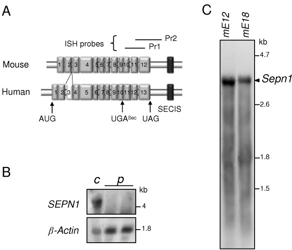
Background: In humans, mutations in the SEPN1 gene, encoding selenoprotein N (SelN), are involved in early onset recessive neuromuscular disorders, referred to as SEPN1-related-myopathies. The mechanisms behind these pathologies are poorly understood since the function of SelN remains elusive. However, previous results obtained in humans and more recently in zebrafish pointed to a potential role for SelN during embryogenesis. Using qRT-PCR, Western blot and whole mount in situ hybridization, we characterized in detail the spatio-temporal expression pattern of the murine Sepn1 gene during development, focusing particularly on skeletal muscles.

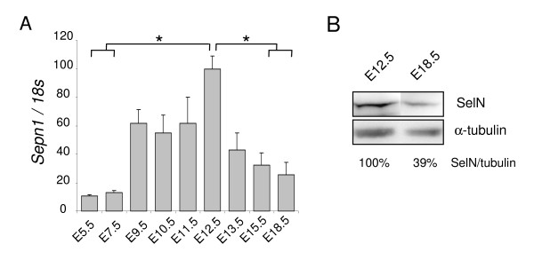
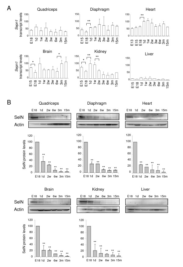
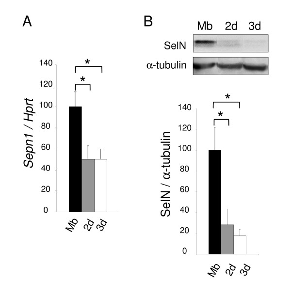
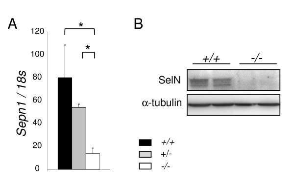
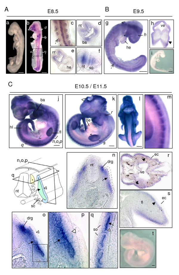
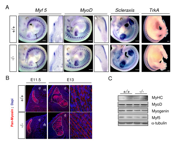
Results: In whole embryos, Sepn1 transcripts were detected as early as E5.5, with expression levels peaking at E12.5, and then strongly decreasing until birth. In isolated tissues, only mild transcriptional variations were observed during development, whereas a striking reduction of the protein expression was detected during the perinatal period. Furthermore, we demonstrated that Sepn1 is expressed early in somites and restricted to the myotome, the sub-ectodermal mesenchyme and the dorsal root ganglia at mid-gestation stages. Interestingly, Sepn1 deficiency did not alter somitogenesis in embryos, suggesting that SelN is dispensable for these processes in mouse.

Conclusion: We characterized for the first time the expression pattern of Sepn1 during mammalian embryogenesis and we demonstrated that its differential expression is most likely dependent on major post-transcriptional regulations. Overall, our data strongly suggest a potential role for selenoprotein N from mid-gestation stages to the perinatal period. Interestingly, its specific expression pattern could be related to the current hypothesis that selenoprotein N may regulate the activity of the ryanodine receptors.


Figures

Figure 1
Figure 1
Sepn1 expression in whole embryos. A: Sepn1 expression was quantified by qRT-PCR on cDNA from whole embryos between E5.5 and E18.5. Normalization is performed on the 18s gene. Expression was detected as early as E5.5 and strongly increased until E12.5. From this stage until birth, a striking decrease of the expression was observed. *, p < 0.05. B: Western blot analysis on 60 μg of proteins from E12.5 and E18.5 embryos was carried out using SelN and α-tubulin (normalization) antibodies. SelN expression was reduced more than two fold at E18.5 compared to E12.5.
Figure 2
Figure 2
Expression of Sepn1 transcript and SelN protein in isolated tissues between late embryonic and post-natal stages. A: Sepn1 expression was quantified by qRT-PCR and normalized to the 18s gene. Expression in quadriceps was quantified from E18 to 15 months; in diaphragm, liver, brain and kidney from E15 to 15 months; in heart from E13 to 15 months. Only mild variations were detected, including between pre- and postnatal stages. Significant variations are indicated for two consecutive points: *, p < 0.05; **, p < 0.01. B: SelN Western blot analyses are shown for quadriceps, diaphragm, heart, liver, brain and kidney at stages E18, postnatal day 1, 2 and 6 weeks, 3 and 15 months. SelN was undetectable in liver. Actin was used as a reference. Quantifications were normalized on the expression measured at E18 and are presented below the blots. For all tissues, a striking reduction of the expression was detected between E18 and postnatal day 1. This decrease was even more marked in the following post-natal stages. Statistical analyses were performed between E18 and all other stages (**, p < 0.01). d: days, w: weeks, m: months.
Figure 3
Figure 3
Sepn1 expression during murine myoblast differentiation. A: Sepn1 qRT-PCR on cDNA from C2C12 myoblasts (Mb) and myotubes after 2 and 3 days (2 d/3 d) in differentiation medium. Hprt served for normalization. A two fold reduction of Sepn1 expression was observed in myotubes compared to undifferentiated cells. B: SelN Western blot analysis on proteins from C2C12 cells (Mb, 2 d, and 3 d), normalized to α-tubulin. Note that the decrease of SelN expression between myoblasts and myotubes is even more marked compared to transcript quantification. *, p < 0.05.
Figure 4
Figure 4
Specificity of the Sepn1 probes. A: Schematic representation of the murine (Sepn1) and human (SEPN1) mRNA. Exons and the SECIS sequence are represented by grey and black boxes, respectively. The human third exon, corresponding to an Alu sequence, is represented by a white box. Target sequences of Pr1 and Pr2 ISH probes are shown above the schema. B: Northern blot analysis, using the Pr1 probe, of RNA extracted from human control fibroblasts (c) and fibroblasts from a RSMD1 patient with a homozygous premature stop codon, leading to SEPN1 mRNA degradation (p). β-actin is used as a reference. A 4.2/4.3 kb band was observed in control fibroblasts but not in RSMD1 fibroblasts, validating the specificity of Pr1 for SEPN1 transcripts. C: Northern blot analysis of RNA from mouse E12 (mE12) and E18 (mE18) whole embryos shows a single 3.4 kb band corresponding to Sepn1 transcript.
Figure 5
Figure 5
SelN deficiency in the murine model. Sepn1 expression analyzed by qRT-PCR (A) and Western blot (B) performed on whole E12.5 littermate embryos: wild-type (+/+), heterozygous (+/-) and homozygous mutants (-/-). 18s gene and α-tubulin were used for normalization. Sepn1 transcript expression was almost abolished in the homozygous mutants and SelN was undetectable by Western blot in these mice. *, p < 0.05.
Figure 6
Figure 6
Sepn1 whole mount in situ hybridization between E8.5 and E11.5. A: E8.5 wild-type embryos hybridized with sense (a) and antisense (b-f) Sepn1 Pr1 probes. No signal was detected with sense probes (a). With the antisense Pr1 probe, all the somites appeared strongly stained (broken arrow - panel c). Sectioning with a vibratome revealed expression also in the branchial arches (d) and the neural tube (f). Note that no signal was observed in the heart (e). B: E9.5 wild-type (g, h) and Sepn1-/- (i) embryos hybridized with the Sepn1 antisense Pr1 probe. Broken arrow in (g) shows stained somites. Mesenchymal staining (black arrowhead) was revealed within vibratome sections (h: section in the forebrain). No signal was seen within the mutant embryos (i). C: E10.5 (j) and E11.5 (k-m) wild-type embryos hybridized with the Sepn1 antisense Pr1 probe. Dorsal region of wild-type embryos is shown in (m). (n-s) High magnification of vibratome sections performed in the trunk (n-q), the head (r), and the limb bud (s). Inset p corresponds to a higher magnification of the outlined region in o. The section plans are indicated in the schema. Sepn1 expression was detected in the sub-ectodermal mesenchyme (black arrowhead), the myotome (arrow) and the dorsal root ganglia (*). Note that overt staining was not observed in the dermomyotome (white arrowhead) nor in the heart. No signal was detected in Sepn1-/- embryos hybridized with Sepn1 antisense probe (t). ba: branchial arches; drg: dorsal root ganglia (*); ec: ectoderm; fl: forelimb; he: heart; hl: hindlimb; nt: neural tube; so: somite; ve: ventricle. Scale bars in (A) = 200 μm, in (B, C) = 400 μm.
Figure 7
Figure 7
Somitogenesis in wild-type and Sepn1-/- embryos. A: Whole mount in situ hybridization, and corresponding vibratome sections, against Myf5, MyoD, Scleraxis (E11.5) and TrkA (E13) performed on Sepn1+/+ and Sepn1-/- embryos. No difference was observed in somite organisation and size (black fragments) between wild-type and mutants embryos. The expression pattern of the tested factors appeared unaltered in absence of SelN. Vibratome sections correspond to the trunk level for Myf5 and to the limb bud for MyoD. B: Immunostaining against myosin heavy chain on E11.5 and E13 wild-type and deficient embryos. The expression patterns were identical between the embryos. Myotome is outlined with a white broken line. C: Western blot analysis against myogenic factors, performed on proteins from wild-type and deficient E12.5 whole embryos, revealed no alteration of their expression in the mutant embryos. α-tubulin was used for normalization. Scale bars = 400 μm. d: drg; nt: neural tube.

References

    1. Allmang C, Krol A. Selenoprotein synthesis: UGA does not end the story. Biochimie. 2006;88:1561–71. doi: 10.1016/j.biochi.2006.04.015. - DOI - PubMed
    1. Papp LV, Lu J, Holmgren A, Khanna KK. From selenium to selenoproteins: synthesis, identity, and their role in human health. Antioxid Redox Signal. 2007;9:775–806. doi: 10.1089/ars.2007.1528. - DOI - PubMed
    1. Kryukov GV, Castellano S, Novoselov SV, Lobanov AV, Zehtab O, Guigo R, Gladyshev VN. Characterization of mammalian selenoproteomes. Science. 2003;300:1439–43. doi: 10.1126/science.1083516. - DOI - PubMed
    1. Jacob C, Giles GI, Giles NM, Sies H. Sulfur and selenium: the role of oxidation state in protein structure and function. Angew Chem Int Ed Engl. 2003;42:4742–58. doi: 10.1002/anie.200300573. - DOI - PubMed
    1. Lu J, Holmgren A. Selenoproteins. J Biol Chem. 2009;284:723–7. doi: 10.1074/jbc.R800045200. - DOI - PubMed

Publication types