Review
. 2009 Nov;28(6):393-422.
doi: 10.1016/j.preteyeres.2009.08.001. Epub 2009 Aug 19.

Aging, age-related macular degeneration, and the response-to-retention of apolipoprotein B-containing lipoproteins

Affiliations
Review

Aging, age-related macular degeneration, and the response-to-retention of apolipoprotein B-containing lipoproteins

Christine A Curcio et al. Prog Retin Eye Res. 2009 Nov.

Abstract

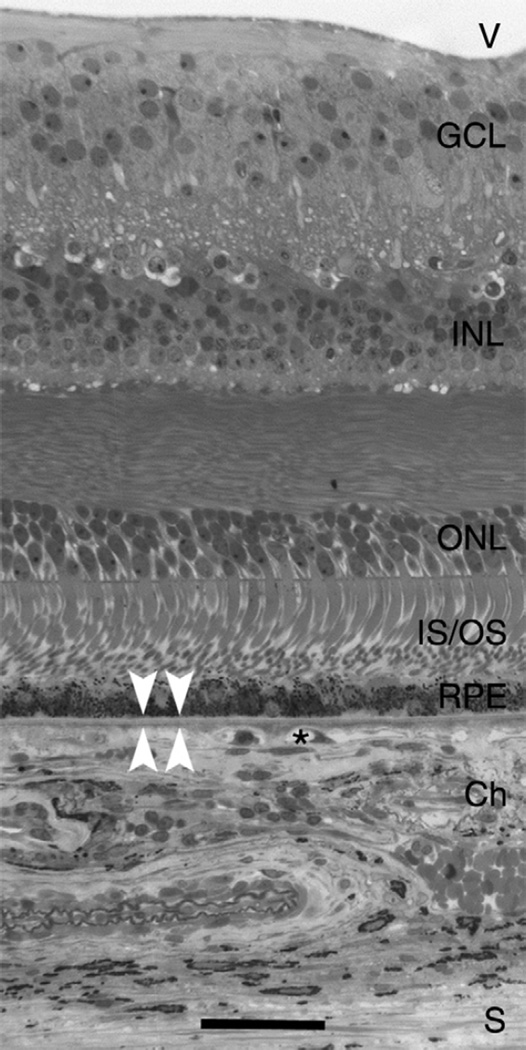
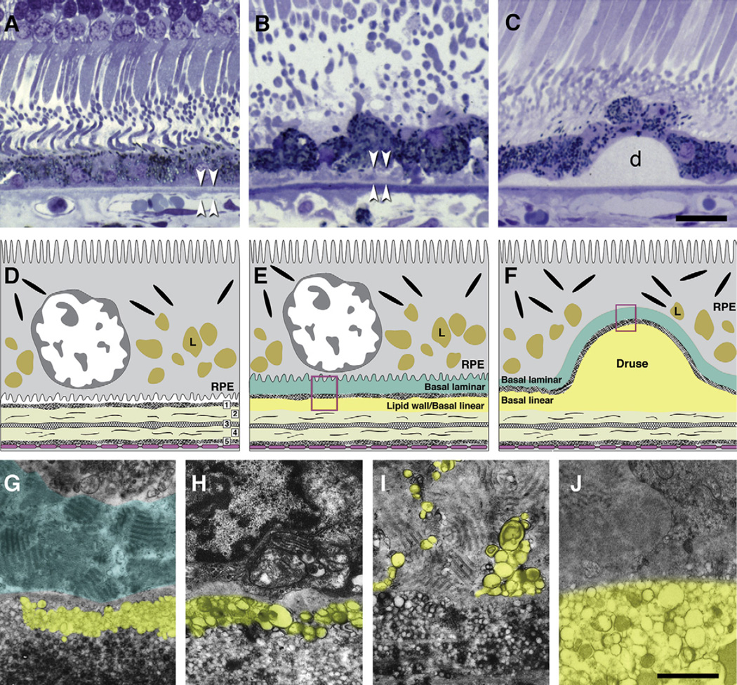
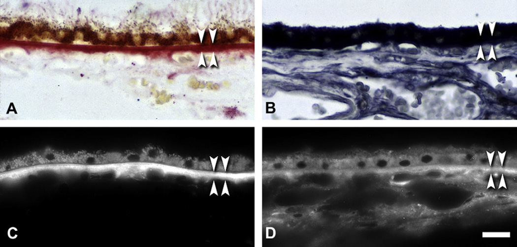
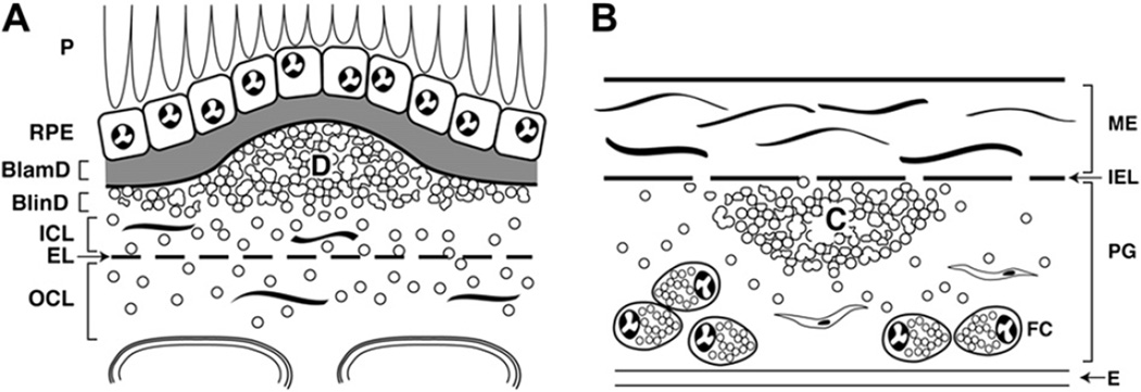
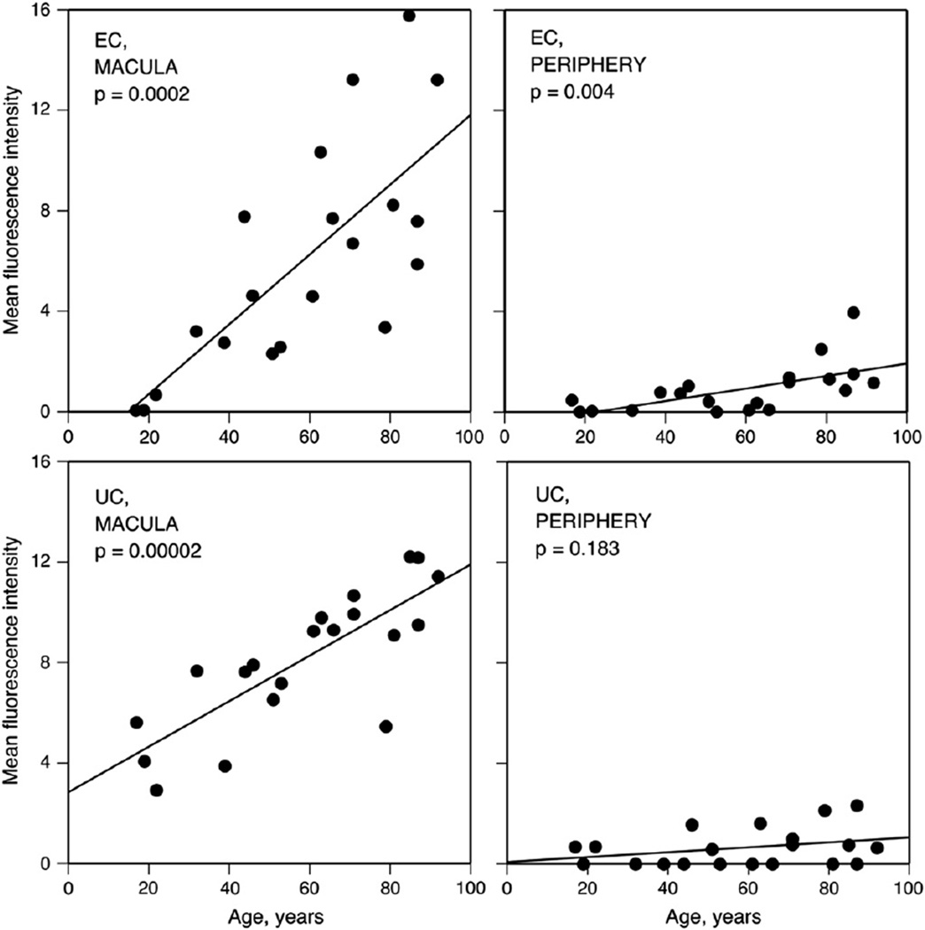
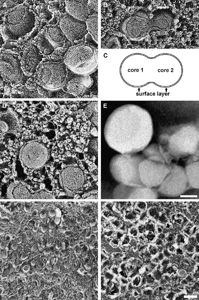
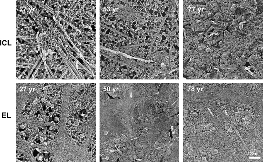
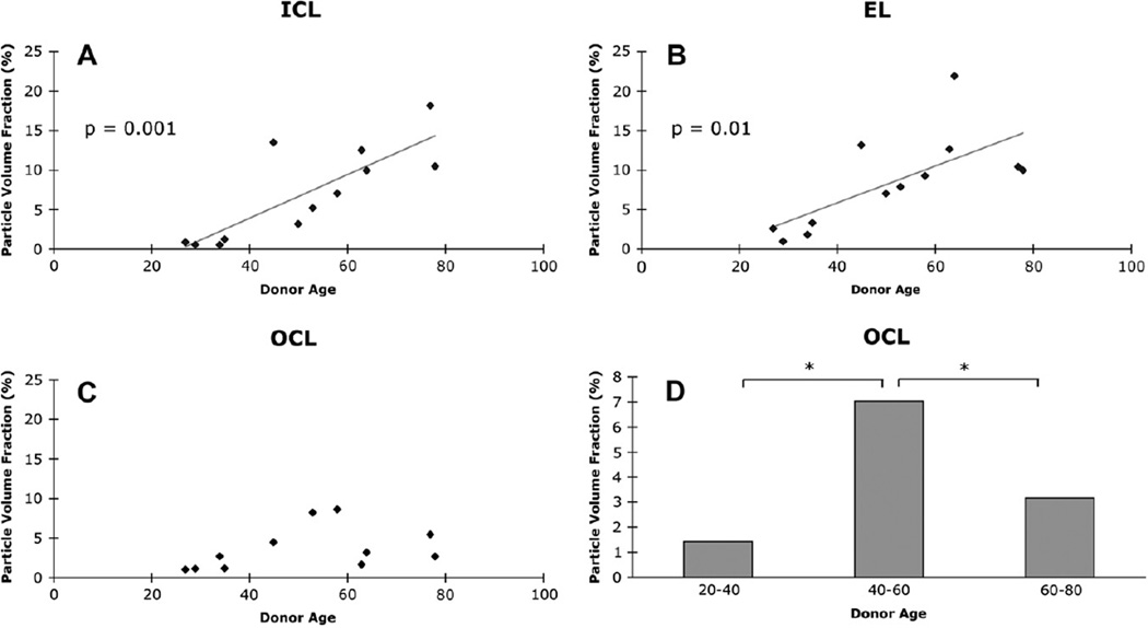
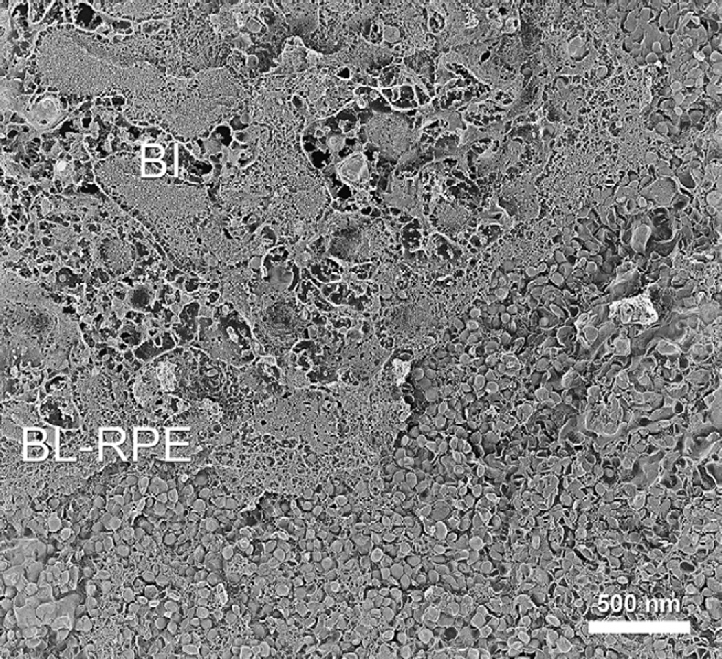
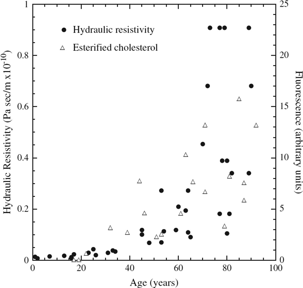
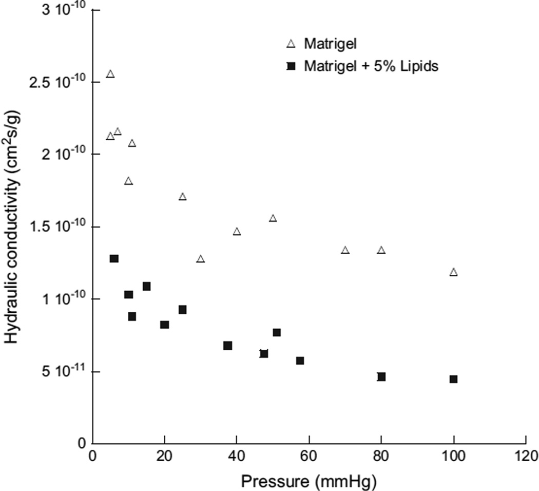
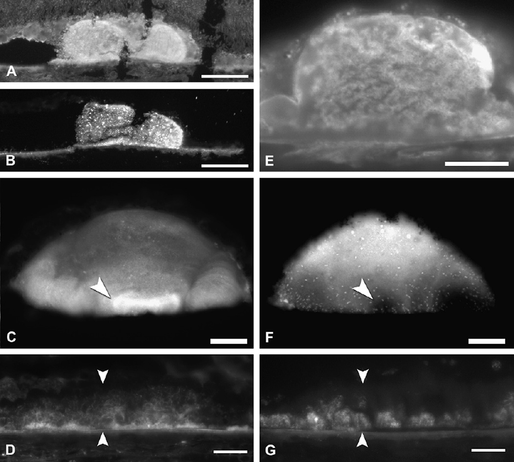
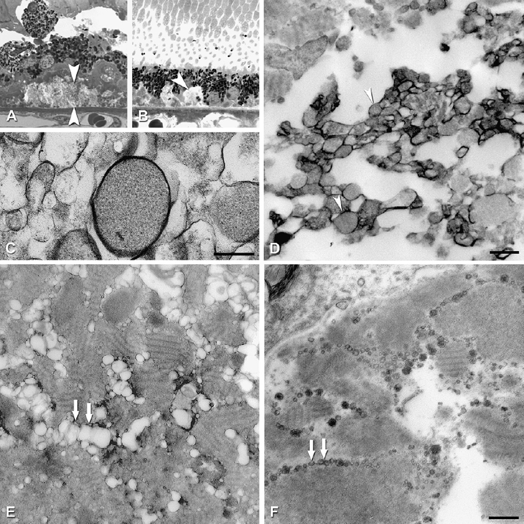
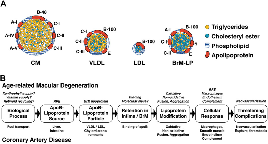
The largest risk factor for age-related macular degeneration (ARMD) is advanced age. A prominent age-related change in the human retina is the accumulation of histochemically detectable neutral lipid in normal Bruch's membrane (BrM) throughout adulthood. This change has the potential to have a major impact on physiology of the retinal pigment epithelium (RPE). It occurs in the same compartment as drusen and basal linear deposit, the pathognomonic extracellular, lipid-containing lesions of ARMD. Here we present evidence from light microscopic histochemistry, ultrastructure, lipid profiling of tissues and isolated lipoproteins, and gene expression analysis that this deposition can be accounted for by esterified cholesterol-rich, apolipoprotein B-containing lipoprotein particles constitutively produced by the RPE. This work collectively allows ARMD lesion formation and its aftermath to be conceptualized as a response to the retention of a sub-endothelial apolipoprotein B lipoprotein, similar to a widely accepted model of atherosclerotic coronary artery disease (CAD) (Tabas et al., 2007). This approach provides a wide knowledge base and sophisticated clinical armamentarium that can be readily exploited for the development of new model systems and the future benefit of ARMD patients.


Figures

Fig. 1
Fig. 1
Chorioretinal anatomy in macula. V, vitreous; GCL, ganglion cell layer; INL, inner nuclear layer; ONL, outer nuclear layer; IS/OS, inner and outer segments of photo-receptors; RPE, retinal pigment epithelium; Ch, choroid; asterisk, choriocapillaris; white arrowheads, BrM; S, sclera. Bar, 50 µm.
Fig. 2
Fig. 2
Bruch’s membrane and ARMD lesions. A–C: 1 µm sections, toluidine blue. Bar in C, 20 µm; A. Normal. RPE, BrM (arrowheads). B. Basal deposits (arrowheads) external to disrupted RPE. C: Druse (d). D–F: cartoons of extracellular lesions. D. BrM has 5 layers in a normal eye: 1, basal lamina of the RPE; 2, inner collagenous layer; 3, elastic layer; 4, outer collagenous layer; 5, basal lamina of the choriocapillary endothelium (fenestrated cells, pink). L, lipofuscin. E. An ARMD eye has basal laminar deposit (BlamD) and basal linear deposit (BlinD) and its precursor, the Lipid Wall. Boxed area is shown in panels G–I. F. Drusen, BlinD, and the Lipid Wall occupy the same plane. Boxed area is shown in panel J. G–J: colorized transmission electron micrographs. Aqua, basal laminar deposit (BlamD); yellow, BlinD, membranous debris, and Lipid Wall. Bar in J, 1 µm G. BlamD (aqua) and Lipid Wall (yellow). H. BlinD. I. Membranous debris crosses BlamD. J. Membranous debris within a large soft druse.
Fig. 3
Fig. 3
Lipid histochemistry of BrM and choroid. Normal macula of a 79 yr old donor. RPE is shown at the top, and choroid at the bottom, of each panel. BrM is bracketed by arrowheads. Bar in D, 20 µm. A. Oil red O stains BrM and lipofuscin. B. Sudan Bromine Black B stains BrM, RPE, and cells throughout choroid. C. Filipin for esterified cholesterol stains BrM. RPE lipofuscin autofluorescence is much less intense than filipin, and it is a different color under ultraviolet illumination. D. Filipin for unesterified cholesterol stains BrM, RPE, and choroidal cells.
Fig. 4
Fig. 4
Cholesterol and lipoprotein basics. A. Cholesterol structure. Cholesterol is esterified to long chain (>16:0) fatty acids at the 3 β-hydroxy group. B. Lipoprotein homeostasis in plasma (with permission (Lusis et al., 2004)). See Section 5.3 for details.
Fig. 5
Fig. 5
ARMD lesions vs plaque. Schematic cross-sections of BrM from an eye with ARMD (B) and atherosclerotic arterial intima (B). Endothelium and vascular lumens (choriocapillary, A; artery, B) are at the bottom. Drawings are not at scale. The thickness of normal BrM and intima is 4–6 µm and 100–300 µm, respectively. Small circles in BrM (A) and PG layer (B) indicate EC-rich lipoproteins, native and modified. A. P = photoreceptors, RPE = retinal pigment epithelium, R-BL = RPE basal lamina, BlamD = basal laminar deposit, BlinD = basal linear deposit, D = druse, ICL = inner collagenous layer EL = elastic layer, OCL = outer collagenous layer. B. ME = musculo-elastic layer, IEL = internal elastic layer, C = lipid-rich core, PG = proteoglycan layer, FC = foam cells. Modified from Malek et al., (2003), with permission from the American Society For Investigative Pathology.
Fig. 6
Fig. 6
BrM cholesterol increases with age. Filipin fluorescence due to EC and UC increases with age in normal BrM. For EC in the macula (A) and periphery (B) and UC in the macula (C) and periphery (D), fluorescence intensity (x 10−6 arbitrary units) for each eye is corrected for background autofluorescence of BrM. Reprinted from (Curcio et al., 2001); copyright is held by Association for Research in Vision and Ophthalmology.
Fig. 7
Fig. 7
BrM lipoproteins, in situ and isolated. A–B, D, F–G. QFDE images of normal BrM (Huang, 2007). E. Negative stain electron microscopy (Li et al., 2005a). A. Core and surface of tightly packed lipoproteins. B. Fused lipoproteins. C. Interpretive schematic based on panel B. D. Small granular particles associated with lipoproteins. E. Isolated particles are large, spherical, and electron-lucent. F,G. Lipoproteins in untreated tissue (F) and in tissue treated with chloroform-methanol to remove lipid (G). Only some surface components, presumably apolipoproteins, remain after extraction. Bar in E = 50 nm, applies to panels A–E. Bar in G = 200 nm, applies to panels F and G.
Fig. 8
Fig. 8
Lipoprotein deposition in BrM layers throughout adulthood. Apparent lipoprotein particle accumulation appears at earlier ages in elastic layer (EL) than inner collagenous layer (ICL). Oblique views of BrM were obtained from normal maculas prepared using QFDE. Bar is 300 nm. Reprinted from (Huang et al., 2007b) with permission from Elsevier.
Fig. 9
Fig. 9
Volume occupied by lipoproteins in BrM layers. Lipoproteins occupy steadily greater volume with age in inner collagenous and elastic layers (ICL, EL) but increase then decrease in outer collagenous layer (OCL). Asterisk indicates significant difference. Reprinted from (Huang et al., 2008a); copyright is held by Association for Research in Vision and Ophthalmology.
Fig. 10
Fig. 10
Lipid Wall. In this oblique view of BrM, lipo proteins are densely packed in the Lipid Wall (lower right corner). Normal macula of an 82-year-old donor, prepared by QFDE. Taken from (Huang et al., 2007b) BI, basal infolding; BL-RPE, basal lamina of the RPE.
Fig. 11
Fig. 11
Hydraulic resistivity and BrM EC in aging. Hydraulic resistivity (Marshall et al., 1998) of excised BrM/choroid (closed symbols) and fluorescence due to histochemically detected esterified cholesterol in sections of normal BrM (Curcio et al., 2001) (open symbols) as a function of age. From (Ethier et al., 2004) Reprinted, with permission, from the Annual Review of Biomedical Engineering, Volume 6 (c)2004 by Annual Reviews www.annualreviews.org.
Fig. 12
Fig. 12
Lipoprotein-derived lipids reduce fluid transport through an artificial matrix. The hydraulicconductivity,asafunctionofperfusionpressure,of1%Matrigel (opentriangle) or 1% Matrigel with 5% LDL added (closed squares). Modified from (McCarty et al., 2008).
Fig. 13
Fig. 13
Cholesterol and apolipoproteins in drusen and deposits. Filipin fluorescence in A–D and F–G. Immunofluorescence in E. Bars in A, B, E 50 µm. Bars in C,D,F,G 20 µm. A. Druse and surrounding chorioretinal tissue contain UC. B. Druse (same as A) and BrM contain EC. Speckles represent lakes of EC. C. A bright UC-rich core (arrowhead) at the base of an isolated, extra-macular druse. D. A thick BlamD (between arrowheads) has bright, delicate fluorescence for UC. E. ApoB immunofluorescence in a druse. F. The core at the base of the same druse as panel C is EC-poor core (arrowhead). This druse also contains EC-rich lakes (speckles). G. A thick BlamD (same as D, between arrowheads) has fluorescence for EC confined to its outer half. Adapted from (Curcio et al., 2005a; Li et al., 2007; Malek et al., 2003).
Fig. 14
Fig. 14
Ultrastructure of cholesterol-containing components of drusen and deposits. Tissue post-fixed with OTAP, from (Curcio et al., 2005b). A. RPE migrating anteriorly and thick BlamD containing cellular processes, bracketed by large arrowheads. B. BlamD containing basal mounds of membranous debris (arrowhead). Bar, 200 nm C. Membranous debris with contents, interior of a large druse. Bar, 200 nm. D. Individual profiles in the basal mound of panel B are solid and surrounded by an electron-dense band. E,F. Bar is 500 nm. E. Linear tracks of partially extracted material resembling membranous debris (double arrows) in BlamD. F. Linear tracks of solid particles (double arrows) in BlamD of the same eye as E, post-fixed with OTAP.
Fig. 15
Fig. 15
Response-to-retention: ARMD, CAD. A. BrM lipoprotein (BrM-LP) compared to other apoB-containing lipoproteins well characterized in plasma. Particle diameters are to scale. Not all the proteins on the BrM lipoprotein are known. B. The hypothesized progression of ARMD has many parallels to the Response-to-Retention hypothesis of atherosclerotic coronary artery disease (Tabas et al., 2007), beginning with apoB-lipoprotein deposition in a vessel wall.

References

    1. A randomized, placebo-controlled, clinical trial of high-dose supplementation with vitamins C and E, beta carotene, and zinc for age-related macular degeneration and vision loss. AREDS Report No. 8. Arch. Ophthalmol. 2001;119:1417–1436. - PMC - PubMed
    1. Executive summary of the third report of the National Cholesterol Education Program (NCEP) expert panel on detection, evaluation, and treatment of high blood cholesterol in adults (Adult Treatment Panel III) JAMA. 2001;285:2486–2497. - PubMed
    1. Adams CWM, Bayliss OB. Lipid histochemistry. In: Glick D, Rosenbaum RM, editors. Techniques of Biochemical and Biophysical Morphology. New York: John Wiley and Sons; 1975. pp. 100–156.
    1. Adiels M, Olofsson SO, Taskinen MR, Borén J. Overproduction of very low-density lipoproteins is the hallmark of the dyslipidemia in the metabolic syndrome. Arterioscler. Thromb. Vasc. Biol. 2008;28:1225–1236. - PubMed
    1. Afshari FT, Fawcett JW. Improving RPE adhesion to Bruch’s membrane. Eye. 2009 - PubMed

Publication types

MeSH terms