Review
. 2009 Nov;23(6):245-55.
doi: 10.1016/j.blre.2009.07.005. Epub 2009 Aug 20.

Transfusion-related acute lung injury (TRALI): current concepts and misconceptions

Affiliations
Review

Transfusion-related acute lung injury (TRALI): current concepts and misconceptions

Christopher C Silliman et al. Blood Rev. 2009 Nov.

Abstract

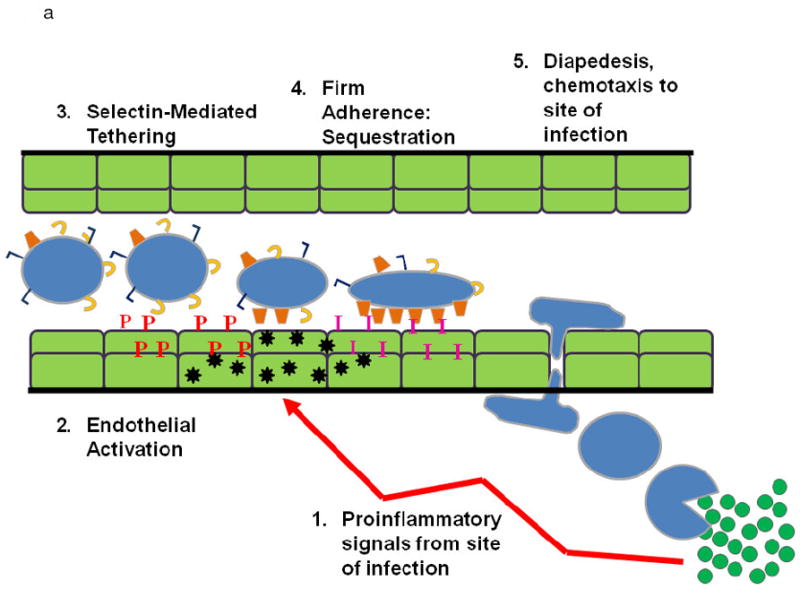
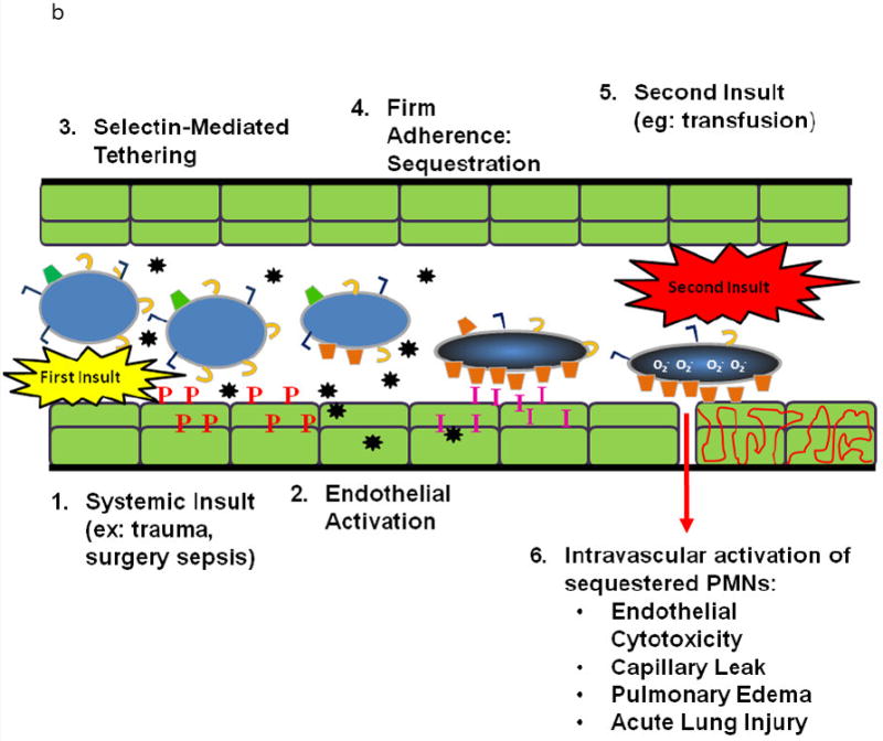
Transfusion-related acute lung injury (TRALI) is the most common cause of serious morbidity and mortality due to hemotherapy. Although the pathogenesis has been related to the infusion of donor antibodies into the recipient, antibody negative TRALI has been reported. Changes in transfusion practices, especially the use of male-only plasma, have decreased the number of antibody-mediated cases and deaths; however, TRALI still occurs. The neutrophil appears to be the effector cell in TRALI and the pathophysiology is centered on neutrophil-mediated endothelial cell cytotoxicity resulting in capillary leak and ALI. This review will detail the pathophysiology of TRALI including recent pre-clinical data, provide insight into newer areas of research, and critically assess current practices to decrease it prevalence and to make transfusion safer.


Figures

Figure 1
Figure 1
A. PMN Physiology: The normal response to infection. From an infection (green circles) in the tissues: (1) pro-inflammatory signals (LPS) are sent out (arrows) that activate ECs (2) causing chemokine synthesis and release (black stars), increases in P-selectin (red P’s) and increased surface expression of intercellular adhesion molecule-1 (ICAM-1, pink I’s). This activation elicits (3) PMN attraction and selectin-mediated tethering (capture) of PMNs between the PMN P=selectin glycoprotein ligand-1 (PSGL-1, tan C’s) and endothelial P-selectin. Capture is then followed by (4) firm PMN CD11b/CD18 (orange trapezoids) : ICAM-1 endothelial cell adherence resulting in PMN pulmonary sequestration. (5) PMNs diapedese through the endothelial cell layer and chemotax to the site of infection and kill the pathogens in the tissues. B. PMN Physiology: PMN-mediated ALI. In response to intravascular stimuli due to a systemic (first) insult, ECs become (1) activated and synthesize and release chemokines (black stars) and increase the surface expression of P-selectin (red P’s) and ICAM-1 (pink I’s) (2). This endothelial activation causes tethering (3). of PMNs via the PMN PSGL-1 (tan C’s) and P-selectin followed by firm adherence (4) via the PMN CD11/CD18 : ICAM-1 from the endothelial surface resulting in pulmonary PMN sequestration. A second intravascular stimulus (insult), transfusion of specific antibodies against host PMN antigens or BRMs activate these primed, adherent PMNs (6) causing release of the microbicidal arsenal from the PMNs ,including O2-, resulting in EC damage, capillary leak, and TRALI.
Figure 1
Figure 1
A. PMN Physiology: The normal response to infection. From an infection (green circles) in the tissues: (1) pro-inflammatory signals (LPS) are sent out (arrows) that activate ECs (2) causing chemokine synthesis and release (black stars), increases in P-selectin (red P’s) and increased surface expression of intercellular adhesion molecule-1 (ICAM-1, pink I’s). This activation elicits (3) PMN attraction and selectin-mediated tethering (capture) of PMNs between the PMN P=selectin glycoprotein ligand-1 (PSGL-1, tan C’s) and endothelial P-selectin. Capture is then followed by (4) firm PMN CD11b/CD18 (orange trapezoids) : ICAM-1 endothelial cell adherence resulting in PMN pulmonary sequestration. (5) PMNs diapedese through the endothelial cell layer and chemotax to the site of infection and kill the pathogens in the tissues. B. PMN Physiology: PMN-mediated ALI. In response to intravascular stimuli due to a systemic (first) insult, ECs become (1) activated and synthesize and release chemokines (black stars) and increase the surface expression of P-selectin (red P’s) and ICAM-1 (pink I’s) (2). This endothelial activation causes tethering (3). of PMNs via the PMN PSGL-1 (tan C’s) and P-selectin followed by firm adherence (4) via the PMN CD11/CD18 : ICAM-1 from the endothelial surface resulting in pulmonary PMN sequestration. A second intravascular stimulus (insult), transfusion of specific antibodies against host PMN antigens or BRMs activate these primed, adherent PMNs (6) causing release of the microbicidal arsenal from the PMNs ,including O2-, resulting in EC damage, capillary leak, and TRALI.
Figure 2
Figure 2
Laboratory investigations into the antibody (Ab) mediated and priming mechanism of TRALI. Patient samples (cellular, serum or plasma) should be as close to the time of the TRALI event as possible to reflect the physiological circumstances of the event. The remains of the transfused blood product provide the most ideal sample for the investigation (primary donor sample), followed by samples from other products manufactured at same collection date from that donor (secondary donor sample) or finally a new sample from the donor (tertiary donor sample). This is because antibody titers and priming/biological response modifiers (BRM) effect can be significantly different between various blood products from the same donor e.g. FFP versus PRBCs, and antibody titers of a donor can vary between blood collected on different dates. Laboratory investigation for Ab mediated TRALI begins with screening for leukocyte antibodies in associated donations and the recipient. Specificities of donor antibodies need to be determined as typing of recipient cells will confirm if there is a cognate Ag on the recipient the donor Ab to interact with. A PMN cross-match (GIFT and GAT) provides in vitro evidence that the donor Ab does react with the recipient’s cells and reveals they type of interaction – agglutination or binding. Primary donor samples should be investigated for their PMN priming activity, especially if leukocyte Abs are not detected.

References

    1. Popovsky MA, Moore SB. Diagnostic and pathogenetic considerations in transfusion-related acute lung injury. Transfusion. 1985 November;25(6):573–7. - PubMed
    1. Kleinman S, Caulfield T, Chan P, Davenport R, McFarland J, McPhedran S, Meade M, Morrison D, Pinsent T, Robillard P, Slinger P. Toward an understanding of transfusion-related acute lung injury: statement of a consensus panel. Transfusion. 2004 December;44(12):1774–89. - PubMed
    1. Toy P, Popovsky MA, Abraham E, Ambruso DR, Holness LG, Kopko PM, McFarland JG, Nathens AB, Silliman CC, Stroncek D. Transfusion-related acute lung injury: definition and review. Crit Care Med. 2005 April;33(4):721–6. - PubMed
    1. Silliman CC, Paterson AJ, Dickey WO, Stroneck DF, Popovsky MA, Caldwell SA, Ambruso DR. The association of biologically active lipids with the development of transfusion-related acute lung injury: a retrospective study. Transfusion. 1997 July;37(7):719–26. - PubMed
    1. Silliman CC, Boshkov LK, Mehdizadehkashi Z, Elzi DJ, Dickey WO, Podlosky L, Clarke G, Ambruso DR. Transfusion-related acute lung injury: epidemiology and a prospective analysis of etiologic factors. Blood. 2003 January 15;101(2):454–62. - PubMed

Publication types