Review
. 2009 Oct;19(5):507-14.
doi: 10.1016/j.sbi.2009.06.003. Epub 2009 Aug 19.

Sialic acids as regulators of molecular and cellular interactions

Affiliations
Review

Sialic acids as regulators of molecular and cellular interactions

Roland Schauer. Curr Opin Struct Biol. 2009 Oct.

Abstract

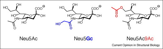
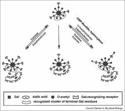
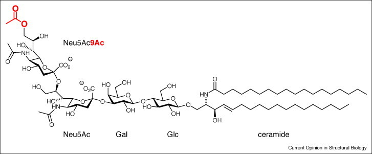
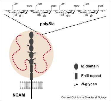
The wide occurrence of sialic acids (Sia) in various chemical forms linked as monomers or polymers in an outstanding position in a multitude of complex carbohydrates of animals and microorganisms renders them as most versatile function modulators in cell biology and pathology. A survey is presented of recent advances in the study of the influences that Sias have as bulky hydrophilic and electronegatively charged monosaccharides on animal cells and on their interaction with microorganisms. Some highlights are: sialylation leads to increased anti-inflammatory activity of IgG antibodies, facilitates the escape of microorganisms from the host's immune system, and in polymeric form is involved in the regulation of embryogenesis and neuronal growth and function. The role of siglecs in immunoregulation, the dynamics of lymphocyte binding to selectins and the interactions of toxins, viruses, and other microorganisms with the host's Sia are now better understood. N-Glycolylneuraminic acid from food is antigenic in man and seems to have pathogenic potential. Sia O-acetylation mediated by various eukaryotic and prokaryotic O-acetyltransferases modulates the affinity of these monosaccharides to mammalian and microbial receptors and hinders apoptosis. The functionally versatile O-acetylated ganglioside GD3 is an onco-fetal antigen.


Figures

Figure 1
Figure 1
The three most frequent sialic acids N-acetylneuraminic acid (Neu5Ac), N-glycolylneuraminic acid (Neu5Gc), and N-acetyl-9-O-acetylneuraminic acid (Neu5,9Ac2), in the figure denominated as Neu5Ac9Ac. Sia are biosynthesized in many steps from glucose leading to both N-acetylmannosamine-6-phosphate (ManNAc-6P) and phosphoenol pyruvate, which are condensed to Neu5Ac-9-phosphate followed by dephosphorylation [6, 7]. The formation of ManNAc-6P from UDP-N-acetylglucosamine (UDP-GlcNAc) is catalyzed by a bifunctional enzyme, the UDP-GlcNAc epimerase/ManNAc kinase (encoded by GNE), which is the key enzyme in Sia biosynthesis and can be feedback-inhibited by CMP-Neu5Ac. After the activation of Neu5Ac with CTP the resulting CMP-Neu5Ac is transported into the Golgi compartment and transferred to nascent glycoconjugates by a large family of monosialyltransferases and polysialyltransferases with varying specificities for glycoproteins and glycolipids [47]. Neu5Ac can be converted to Neu5Gc on the CMP-Neu5Ac level in the cytosol by the action of CMP-Neu5Ac hydroxylase (CMAH) [6]. The activity of this enzyme was lost during human evolution [38]. Enzymes O-acetylating Sia at C-4 of the pyranose ring or at C-7 to C-9 of the side-chain also seem to be frequent, studied best in animals in bovine submandibular gland [6, 17, 48]. Only prokaryotic sialate-O-acetyltransferase genes could be identified so far and some insight into the catalytic mechanism of OatC from Neisseria meningitidis was obtained [49]. Hydroxylation and O-acetylation as well as other natural or artificial Sia modifications strongly influence the physiological properties of Sia.
Figure 2
Figure 2
Model of the association and dissociation of cells regulated by the loss, restoration, and modification of Sia by the action of sialidases, sialyltransferases, sialate-O-acetyltransferases, CMP-Neu5Ac hydroxylase, and sialate-O-acetylesterases. Only after the unmasking of a sufficient number of galactose residues (a ‘cluster’), attachment to another cell, for example a macrophage, via galactose-recognizing receptors is possible. O-Acetyl (black dot) or N-glycolyl (black triangle) groups may inhibit the enzymatic release of Sia and the interaction of these monosaccharides with Sia-recognizing receptors, for example, siglecs, or with viruses and bacteria. These Sia modifications are involved in fine-tuning of recognition events or can even be considered as molecular switches. O-Acetylation may fortify masking of immunogenic epitopes on microorganisms and enable escape from immune defense [49]. In this model symbols for Sia-recognizing receptors have not been drawn, and the symbol used for the Sia molecule could also represent oligosialic acids or polysialic acids, which may carry non-natural N-acyl or O-acyl modifications. The degradation of Sia is initiated by sialidases [6, 50, 51], which strongly influence the behavior of normal and malignant cells. Polysialic acids can be split into Sia oligosaccharides by endosialidases from phages [52]. For de-O-acetylation of Sia a variety of esterases from animals and microorganisms are known [6, 53].
Figure 3
Figure 3
Structure of 9-O-acetylated disialo-ganglioside GD3 (9-OAcGD3; Neu5,9Ac2-α2,8Neu5Acα2,3Galβ1,4Glcβ1,1ceramide). The terminal Sia carries an O-acetyl (red) at C-9, which renders this frequent ganglioside an important regulator and biomarker of growth, differentiation, and malignancy of neuro-ectodermal tissues, as well as of lymphocyte function and apoptosis [17]. GD3 can directly be O-acetylated at C-7 by mammalian sialate-O-acetyltransferases, followed by migration of the O-acetyl to C-9. Therefore, both 7-OAcGD3 and 9-OAcGD3 occur in lymphocytes and tissues in various amounts.
Figure 4
Figure 4
Polysialylation of the neuronal cell-aggregation molecule NCAM of mouse (modified from Mühlenhoff et al. [19]). The extracellular part of NCAM consists of five immunoglobulin (Ig)-like domains and two fibronectin type III (FnIII) repeats. Various isoforms exist, which are either attached to the plasma membrane by a glycosylphosphatidylinositol (GPI) anchor or are inserted into the membrane by trans-membrane protein domains. NCAM has six N-glycosylation sites, as indicated. In PSA–NCAM the inner (5th and 6th) N-glycans carry one or more PSA chains with up to 90 α2,8-linked Sia residues (red dots). The pink-shaped sphere represents the hydrodynamic radius of PSA.

Similar articles

Cited by

References

    1. Kamerling J.P., Boons G.-J., Lee Y.C., Suzuki A., Taniguchi N., Voragen A.G.J., editors. vols. 1–4. Elsevier; Oxford: 2007. (Comprehensive Glycoscience—From Chemistry to Systems Biology).
    2. This multiauthor, well-edited book series comprises all aspects of structure, analysis, chemical and biochemical synthesis, catabolism, molecular biology and cell biological, and pathophysiological aspects of complex carbohydrates. The reader's attention is drawn to the following chapters: Kobata A: Glycoprotein glycan structures, vol. 1, 39–72; Yu RK, Yanagisawa M, Ariga T: Glycosphingolipid structures, vol. 1, 73–122; Miyagi T, Yamaguchi K: Sialic acids, vol. 3, 297–323.

    1. Royle L., Matthews E., Corfield A., Berry M., Rudd P.M., Dwek R.A., Carrington S.D. Glycan structures of ocular surface mucins in man, rabbit and dog display species differences. Glycoconj J. 2008;25:763–773. - PubMed
    1. Varki A. Sialic acids in human health and disease. Trends Mol Med. 2008;14:351–360. - PMC - PubMed
    1. Janas T., Janas T. Polysialic acids: structure and properties. In: Dumitriu S., editor. Polysaccharides — Structural Diversity and Functional Versatility. edn 2. Marcel Dekker; New York: 2005. pp. 707–727.
    1. von Itzstein M., Thomson R. Anti-influenza drugs: the development of sialidase inhibitors. In: Kräusslich H.-G., Bartenschlager R., editors. vol. 189. Springer-Verlag; Berlin, Heidelberg: 2009. pp. 111–154. (Antiviral Strategies. Handbook Exp Pharmacol). - PubMed

LinkOut - more resources