Review
. 2009 Oct;13(4):451-9.
doi: 10.1016/j.cbpa.2009.07.018. Epub 2009 Aug 19.

Structures and mechanisms of the mycothiol biosynthetic enzymes

Affiliations
Review

Structures and mechanisms of the mycothiol biosynthetic enzymes

Fan Fan et al. Curr Opin Chem Biol. 2009 Oct.

Abstract

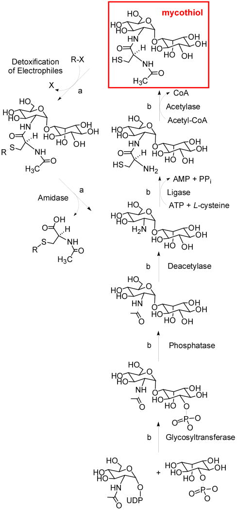
In the past decade, the genes encoding all four enzymes responsible for the biosynthesis of mycothiol in Mycobacterium tuberculosis have been identified. Orthologs of each of these have been stably expressed and structurally characterized. The chemical mechanisms of all the four have also been studied. Because of the unique phylogenetic distribution of mycothiol, and the enzymes responsible for its biosynthesis, these enzymes represent interesting potential targets for antimycobacterial agents.


Conflict of interest statement

Ethnic in Publishing, General Statement: The authors declare that they have no competing financial interests.

Figures

Figure 1
Figure 1
A) Three-dimensional structure of MshA monomer (PDBID 3c4v). B) Stereo stick diagram of ternary complex model. 1l-Ins-1-P carbons colored yellow, UDP-GlcNAc carbons colored green. Important hydrogen bonding interactions shown as grey dotted lines, and the interaction of the 3-OH of 1l-Ins-1-P with the UDP-GlcNAc at the site of reaction chemistry shown as cyan dotted lines. C) Proposed SNi – substrate assisted catalysis mechanism, featuring oxycarbenium-ion like transition state, with asymmetric phosphor bond breakage, and glycosidic bond formation.
Figure 2
Figure 2
A) Three-dimensional structure of MshB monomer (PDBID 1q74). B) Stereo stick diagram enzyme active site of MshB, with the catalytically relevant zinc atom, coordinated by His13, Asp16 and His146 along with the two water molecules. C) Proposed catalytic mechanism for MshB. In this mechanism, the highly conserved Asp15 acts as general base to deprotonate the active site water molecule, which in turn carries out a nucleophilic attack to the amide carbonyl polarized by the zinc atom.
Figure 3
Figure 3
A) Three-dimensional structure of MshC monomer (PDBID 3c8z), resolved at 1.6 Å. B) A diagram illustrating the active site of MshC, in presence of tight binding inhibitor CSA. C) The proposed Bi Uni Uni Bi Ping Pong kinetic mechanism of MshC, based on steady state kinetic as well as PIX studies.
Figure 4
Figure 4
Structure and mechanism of MshD. A) Ribbon diagram of the CoA-DAM-MshD ternary complex. Ligands shown as sticks colored by atom type. B) DYNDOM analysis diagram showing the domain movements upon formation of the ternary complex. The MshD-AcCoA complex (open; PDBID 1ozp) shown as green trace, while the MshD-CoA-DAM ternary complex (closed; PDBID 2c27) shown with the N-terminal domain in cyan and the C-terminal domain in blue. The hinge residues are colored red, and the orange arrow is the axis of the domain rotation. α2 is colored magenta in both the open and closed conformation as a reference point. The ligands for the MshD-AcCoA complex and the AcCoA bound to the N-terminal domain are omitted for clarity. C) Direct attack of the desacetylmycothiol amine upon the acetyl group of AcCoA. Chemistry is facilitated by the partial charge of the backbone amide of Leu238 stabilizing the tetrahedral intermediate and Glu324 and Tyr294 acting as active site base and acid, respectively.
Scheme 1
Scheme 1
Detoxification mechanism (path a) and biosynthetic pathway of MSH (path b)

References

    1. Newton GL, Unson MD, Anderberg SJ, Aguilera JA, Oh NN, delCardayre SB, Av-Gay Y, Fahey RC. Characterization of Mycobacterium smegmatis mutants defective in 1-d-myo-inosityl-2-amino-2-deoxy-alpha-d-glucopyranoside and mycothiol biosynthesis. Biochem Biophys Res Commun. 1999;255:239–244. - PubMed
    1. Newton GL, Arnold K, Price MS, Sherrill C, Delcardayre SB, Aharonowitz Y, Cohen G, Davies J, Fahey RC, Davis C. Distribution of thiols in microorganisms: mycothiol is a major thiol in most actinomycetes. J Bacteriol. 1996;178:1990–1995. - PMC - PubMed
    1. Newton GL, Av-Gay Y, Fahey RC. A novel mycothiol-dependent detoxification pathway in mycobacteria involving mycothiol S-conjugate amidase. Biochemistry. 2000;39:10739–10746. - PubMed
    1. Rawat M, Newton GL, Ko M, Martinez GJ, Fahey RC, Av-Gay Y. Mycothiol-deficient Mycobacterium smegmatis mutants are hypersensitive to alkylating agents, free radicals, and antibiotics. Antimicrob Agents Chemother. 2002;46:3348–3355. - PMC - PubMed
    2. With one chemical mutant and two transposon mutants, the authors demonstrated that MshC is important for the sensitivity of Mycobacteria smegmatis towards a variety of stresses, including the suseptibility of these strains to commonly used antibiotics, erythromycin, azithromycin, vancomycin, penicillin G, rifamycin and rifampin.

    1. Park JH, Roe JH. Mycothiol regulates and is regulated by a thiol-specific antisigma factor RsrA and sigma(R) in Streptomyces coelicolor. Mol Microbiol. 2008;68:861–870. - PubMed

Publication types

MeSH terms

LinkOut - more resources