. 2009 Sep 4;5(3):290-7.
doi: 10.1016/j.stem.2009.06.003. Epub 2009 Aug 20.

The Drosophila hindgut lacks constitutively active adult stem cells but proliferates in response to tissue damage

Affiliations

The Drosophila hindgut lacks constitutively active adult stem cells but proliferates in response to tissue damage

Donald T Fox et al. Cell Stem Cell. .

Abstract

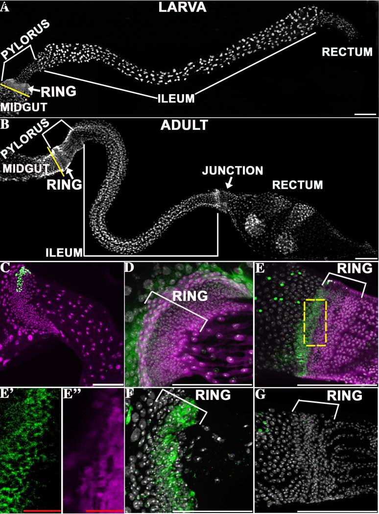
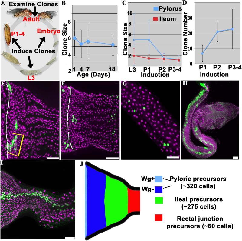
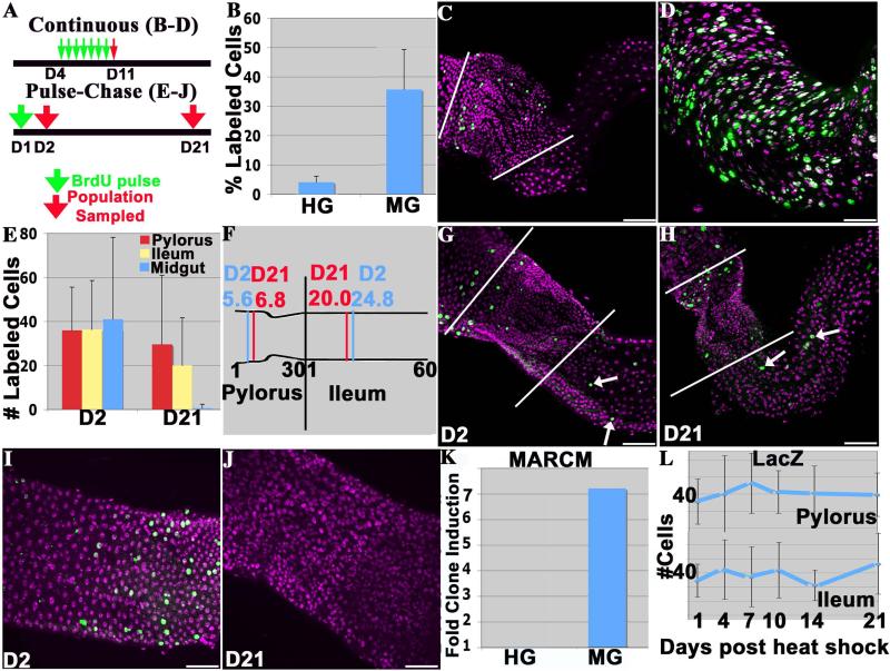
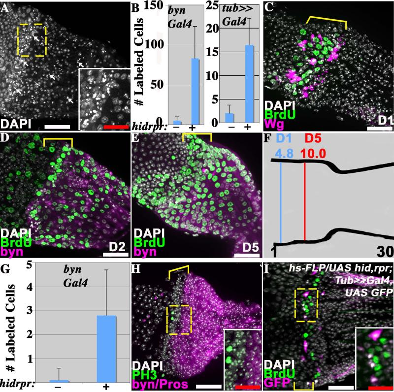
The adult Drosophila hindgut was recently reported to contain active, tissue-replenishing stem cells, like those of the midgut, but located within an anterior ring so as to comprise a single giant crypt. In contrast to this view, we observed no active stem cells and little cell turnover in adult hindgut tissue based on clonal marking and BrdU incorporation studies. Again contradicting the previous proposal, we showed that the adult hindgut is not generated by anterior stem cells during larval/pupal development. However, severe tissue damage within the hindgut elicits cell proliferation within a ring of putative quiescent stem cells at the anterior of the pylorus. Thus, the hindgut does not provide a model of tissue maintenance by constitutively active stem cells, but has great potential to illuminate mechanisms of stress-induced tissue repair.


Figures

Figure 1
Figure 1. Similar larval and adult hindgut anatomy
The general structure of the larval hindgut (A) and the adult hindgut (B) is shown. The major hindgut subdivisions and the pyloric rings are indicated. The yellow line denotes the midgut/hindgut boundary. C) A representative lacZ clone (green) induced in the first instar and analyzed in L3. D, E) Wg protein (green, cytoplasmic) is highly expressed in 1-3 rows of anterior ring cells in the pylorus of both larvae (D) and adults (E). Prospero-positive enteroendocrine cells (green, nuclear in E) mark the midgut, which lies anterior to the ring. More posterior ring cells strongly express byn-Gal4::UAS-GFP (purple). The separate channels are shown for a section indicated by the dashed box in E’ (Wg, green) and E” (byn-GAL4::UAS-GFP, purple). F) Posterior larval ring cells express the Notch activation reporter GBE-Su(H) lacZ (green). G) The adult ring, in contrast, does not express GBE-Su(H) lacZ (green). All panels- anterior to left, DAPI= nuclei (white). Scale bars- white=100μm, red=12.5μm.
Figure 2
Figure 2. Separate progenitors produce the adult pylorus and ileum
A) The clone induction scheme used to study adult hindgut progenitors. B) The size of pyloric clones induced during pupal development does not change significantly during adulthood. C) Pyloric (blue) and ileal (red) clones induced during late larval development (L3) are small, and decline further in size when induced after pupal day 1 (P1). D) The number of pyloric clones per gut induced by a standard heat shock increases during pupal development. E-G) Examples of clones induced in late larvae or pupae and analyzed in adults. E) Adult pylorus showing an L3 pyloric clone (yellow bracket) in contact with the posterior boundary of the Wg ring (dashed line). F) Adult pylorus showing 6 P1 pyloric clones, 4 of which do not contact the Wg ring (boundary = dashed line). G) P1 ileal clones, showing small size (1-2 cells). H) Rare large spontaneous clone spanning pylorus and ileum. I) Example of large lacZ clone induced during embryogenesis. J) Proposed fate map of the larval pylorus. All images- anterior to the left. DAPI (purple), clones (green) were labeled with membrane GFP (E,H) or nuclear lacZ (F,G,I). N > 70 clones/time point. Scale bars = 25μm. Lifecycle image adapted from http://flymove.uni-muenster.de/
Figure 3
Figure 3. The adult hindgut is not maintained by active stem cells
A) Experimental plan for continuous or pulse chase BrdU labeling studies; green arrow = BrdU addition, red arrow = tissue analysis. B) Percentage of BrdU-positive cells (average +/− standard deviation) in hindgut (HG) and midgut (MG) after seven days of BrdU exposure. C) BrdU incorporation following continuous labeling in the hindgut appears sporadic and is limited to the pylorus. D) Widespread BrdU incorporation in the midguts of the same animals. E) Slow turnover of BrdU-labeled cells in hindguts compared to the midguts of the same animals following the pulse-chase protocol. N >23 guts/time point. F) Absence of significant posterior cell movement over a 20-day period. The mean cell position of BrdU positive cells along the proximal-distal axes of the pylorus (30 cell diameters long) or the ileum (60 cell diameters long) at day 2 vs day 21. N > 200 pyloric and >100 ileal cells/time point. G) A hindgut 1d after the BrdU pulse (D2), or H) 20d post-pulse (D21). Arrows- ileal labeling; white lines= pyloric boundary. I) A midgut 1d after the BrdU pulse (D2), or J) 20d post-pulse (D21). K) The increase in the number of MARCM clones 21d after adult heat-shock, normalized to the number of clones in non-heat shocked controls. No increase is seen in the hindgut, unlike the stem cell-based midgut where the number of clones is strongly increased. L) The number of lacZ clones induced in 4-day-old adults per gut is stable during adulthood. 12-34 animals were scored at each time point. All images- anterior to the left. DAPI (purple), BrdU (green), Scale bar= 50μm.
Figure 4
Figure 4. The pylorus proliferates in response to damage
In A-H) adults of the genotype UAS hid rpr ; Gal80ts ; byn-GAL4 UAS-GFP were shifted to 30 °C to induce damage in the hindgut. A) Punctate nuclei of dying pyloric cells (arrows) 2 days after damage induction. B) The number of BrdU-labeled adult hindgut cells (average +/− standard deviation) is greatly increased following damage induction (+), relative to controls lacking hid and rpr expression (−). Note: A Tub-GAL4 FLP-out system was substituted for byn-GAL4 in one experiment (see I). C). BrdU incorporation occurs preferentially in the adult pyloric Wg ring (bracket) one day after damage induction. D) BrdU incorporation in the adult pylorus 2 days after damage induction is seen both in the Wg ring (bracket) and more posteriorly. E) Adult pyloric cells shifted to the non-permissive temperature and labeled with BrdU for one day (as in C) were examined after 4 additional days at 18 °C. Labeled cells are now seen throughout the pylorus. Bracket = Wg ring. F) The mean position of BrdU-labeled cells after 1 day of damage (D1, see panel C) and after 5 days (D5, see panel E) demonstrates cell movement. G) Cells positive for phosphohistone H3 (PH3) (average +/− standard deviation) are greatly increased in adult hindguts following damage induction (+), relative to controls lacking hid and rpr expression (−). H) Cell division marked by PH3 (green) two days after damage induction is confined to the Wg ring (bracket). I) BrdU incorporation 1 day after the induction of FLP-out clones marked with GFP (pink) and expressing hid and rpr (full genotype indicated on panel). BrdU incorporation (green) is observed within the Wg ring (bracket) in unrecombined cells adjacent to GFP-expressing cells (see inset). All images- anterior to the left. Dashed box = region shown in inset. In D, E and H, byn = GFP (purple); in I, GFP = UAS hid,rpr positive. Pros=Prospero, Wg = Wingless. Scale bars- white=25μm, red=12.5μm.

Comment in

References

    1. Ahmad K, Golic K. Somatic reversion of chromosomal position effects in Drosophila melanogaster. Genetics. 1996;144:657–660. - PMC - PubMed
    1. Amcheslavsky A, Jiang J, Ip YT. Tissue damage-induced intestinal stem cell division in Drosophila. Cell Stem Cell. 2009;4:49–61. - PMC - PubMed
    1. Buchon N, Broderick NA, Poidevin M, Pradervand S, Lemaitre B. Drosophila intestinal response to bacterial infection: activation of host defense and stem cell proliferation. Cell host & microbe. 2009;5:200–211. - PubMed
    1. Casali A, Batlle E. Intestinal Stem Cells in Mammals and Drosophila. Cell Stem Cell. 2009;4:124–127. - PubMed
    1. Dor Y, Brown J, Martinez OI, Melton DA. Adult pancreatic beta-cells are formed by self-duplication rather than stem-cell differentiation. Nature. 2004;429:41–46. - PubMed

Publication types

Substances

LinkOut - more resources