

Persistence of DNA damage following exposure of human bladder cells to chronic monomethylarsonous acid
- PMID: 19699219
- PMCID: PMC2783778
- DOI: 10.1016/j.taap.2009.08.016
Persistence of DNA damage following exposure of human bladder cells to chronic monomethylarsonous acid
Abstract
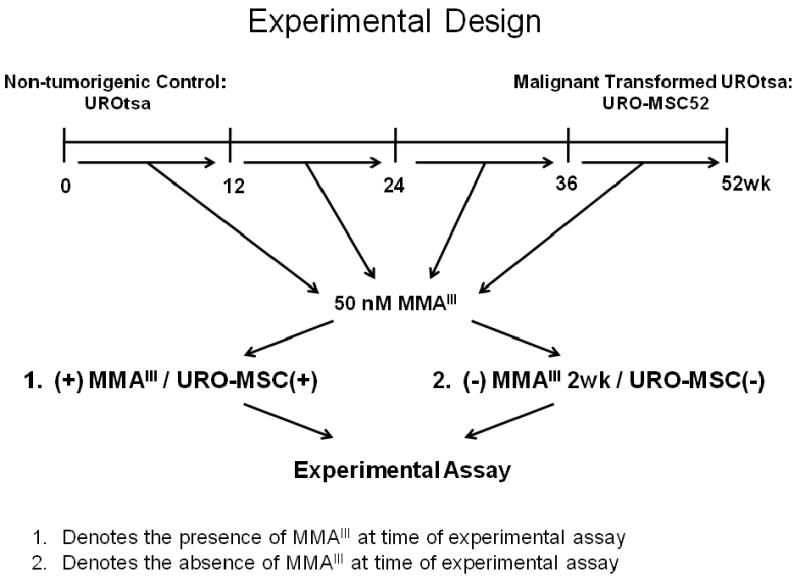
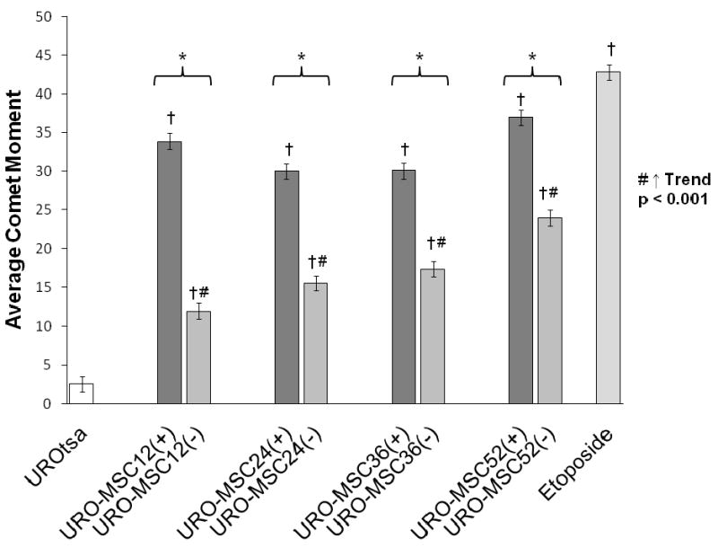
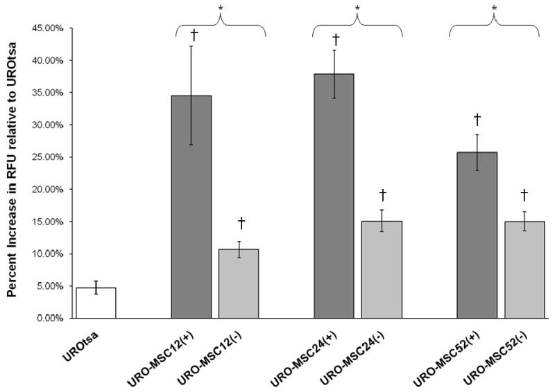
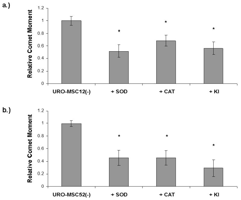
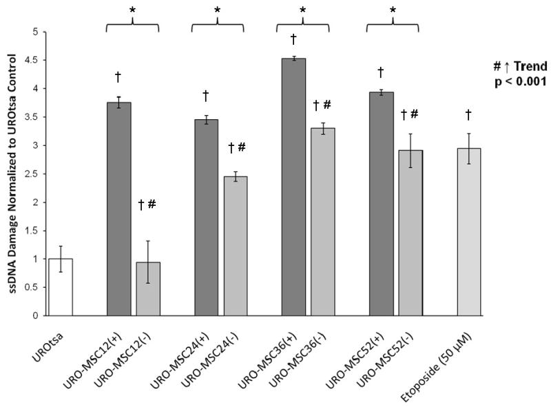
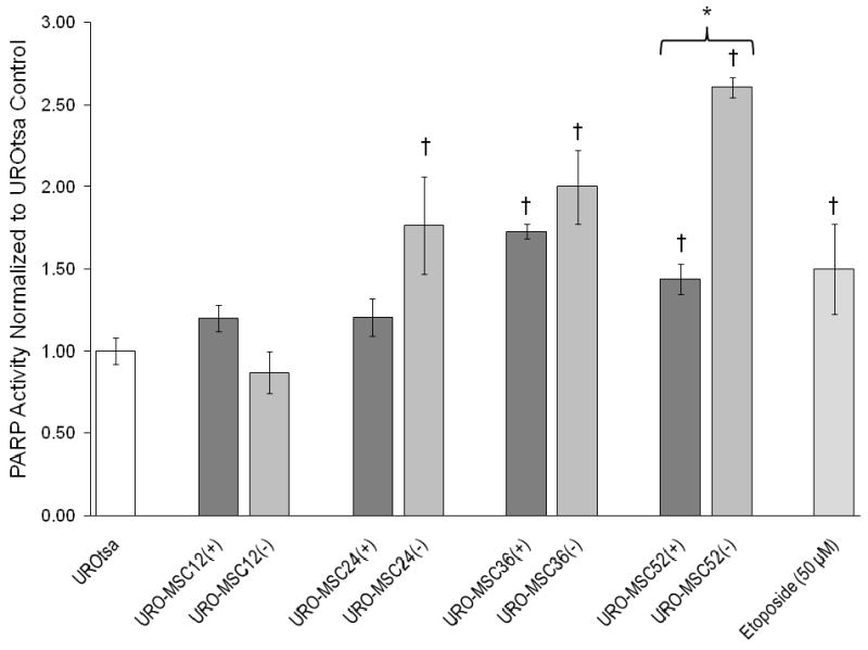
Malignant transformation was demonstrated in UROtsa cells following 52-weeks of exposure to 50 nM monomethylarsonous acid (MMA(III)); the result was the malignantly transformed cell line, URO-MSC. URO-MSC cells were used to study the induction of DNA damage and the alteration of DNA repair enzymes in both the presence of MMA(III) [URO-MSC(+)] and after subsequent removal of MMA(III) [URO-MSC(-)] following chronic, low-level exposure. In the presence of MMA(III), URO-MSC(+) cells demonstrated a sustained increase in DNA damage following 12-weeks of exposure; in particular, a significant increase in DNA single-strand breaks at 12-weeks of exposure consistently elevated through 52 weeks. The persistence of DNA damage in URO-MSC cells was assessed after a 2-week removal of MMA(III). URO-MSC(-) cells demonstrated a decrease in DNA damage compared to URO-MSC(+); however, DNA damage in URO-MSC(-) remained significantly elevated when compared to untreated UROtsa and increased in a time-dependent manner. Reactive oxygen species (ROS) were demonstrated to be a critical component in the generation of DNA damage determined through the incubation of ROS scavengers with URO-MSC cells. Poly (ADP-ribose) polymerase (PARP) is a key repair enzyme in DNA single-strand break repair. URO-MSC(+) resulted in a slight increase in PARP activity after 36-weeks of MMA(III) exposure, suggesting the presence of MMA(III) is inhibiting the increase in PARP activity. In support, PARP activity in URO-MSC(-) increased significantly, coinciding with a subsequent decrease in DNA damage demonstrated in URO-MSC(-) compared to URO-MSC(+). These data demonstrate that chronic, low-level exposure of UROtsa cells to 50 nM MMA(III) results in: the induction of DNA damage that remains elevated upon removal of MMA(III); increased levels of ROS that play a role in MMA(III) induced-DNA damage; and decreased PARP activity in the presence of MMA(III).
Figures
Similar articles
-
Interdependent genotoxic mechanisms of monomethylarsonous acid: role of ROS-induced DNA damage and poly(ADP-ribose) polymerase-1 inhibition in the malignant transformation of urothelial cells.Toxicol Appl Pharmacol. 2011 Nov 15;257(1):1-13. doi: 10.1016/j.taap.2011.08.029. Epub 2011 Sep 10. Toxicol Appl Pharmacol. 2011. PMID: 21925530 Free PMC article.
-
Monomethylarsonous acid induces transformation of human bladder cells.Toxicol Appl Pharmacol. 2006 Oct 1;216(1):69-79. doi: 10.1016/j.taap.2006.04.011. Epub 2006 Jun 27. Toxicol Appl Pharmacol. 2006. PMID: 16806342 Free PMC article.
-
Arsenite and monomethylarsonous acid generate oxidative stress response in human bladder cell culture.Toxicol Appl Pharmacol. 2006 Nov 15;217(1):7-14. doi: 10.1016/j.taap.2006.07.004. Epub 2006 Jul 21. Toxicol Appl Pharmacol. 2006. PMID: 16930658
-
Immortalized human urothelial cells as a model of arsenic-induced bladder cancer.Toxicology. 2008 Jun 27;248(2-3):67-76. doi: 10.1016/j.tox.2008.03.020. Epub 2008 Mar 30. Toxicology. 2008. PMID: 18456381 Review.
-
Modulating poly (ADP-ribose) polymerase activity: potential for the prevention and therapy of pathogenic situations involving DNA damage and oxidative stress.Curr Pharm Biotechnol. 2002 Sep;3(3):275-83. doi: 10.2174/1389201023378265. Curr Pharm Biotechnol. 2002. PMID: 12164482 Review.
Cited by
-
Interdependent genotoxic mechanisms of monomethylarsonous acid: role of ROS-induced DNA damage and poly(ADP-ribose) polymerase-1 inhibition in the malignant transformation of urothelial cells.Toxicol Appl Pharmacol. 2011 Nov 15;257(1):1-13. doi: 10.1016/j.taap.2011.08.029. Epub 2011 Sep 10. Toxicol Appl Pharmacol. 2011. PMID: 21925530 Free PMC article.
-
Expression Of Selected Pathway-Marker Genes In Human Urothelial Cells Exposed Chronically To A Non-Cytotoxic Concentration Of Monomethylarsonous Acid.Toxicol Rep. 2014;1:421-434. doi: 10.1016/j.toxrep.2014.07.004. Toxicol Rep. 2014. PMID: 25177542 Free PMC article.
-
Risk assessment of small organoarsenic species in food.EFSA J. 2024 Jul 2;22(7):e8844. doi: 10.2903/j.efsa.2024.8844. eCollection 2024 Jul. EFSA J. 2024. PMID: 38957748 Free PMC article.
-
Global gene expression changes in human urothelial cells exposed to low-level monomethylarsonous acid.Toxicology. 2012 Jan 27;291(1-3):102-12. doi: 10.1016/j.tox.2011.11.002. Epub 2011 Nov 17. Toxicology. 2012. PMID: 22108045 Free PMC article.
-
Low level exposure to monomethyl arsonous acid-induced the over-production of inflammation-related cytokines and the activation of cell signals associated with tumor progression in a urothelial cell model.Toxicol Appl Pharmacol. 2010 Apr 15;244(2):162-73. doi: 10.1016/j.taap.2009.12.029. Epub 2010 Jan 4. Toxicol Appl Pharmacol. 2010. PMID: 20045430 Free PMC article.
References
-
- Ahlborn GJ, Nelson GM, Ward WO, Knapp G, Allen JW, Ouyang M, Roop BC, Chen Y, O'Brien T, Kitchin KT, Delker DA. Dose response evaluation of gene expression profiles in the skin of K6/ODC mice exposed to sodium arsenite. Toxicol Appl Pharmacol. 2008;227:400–416. - PubMed
-
- Aposhian HV. Enzymatic methylation of arsenic species and other new approaches to arsenic toxicity. Annu Rev Pharmacol Toxicol. 1997;37:397–419. - PubMed
-
- Aposhian HV, Gurzau ES, Le XC, Gurzau A, Healy SM, Lu X, Ma M, Yip Li, Zakharyan RA, Maiorina RM, Dart RC, Tircus MG, Gonzalez-Ramirez D, Morgan DL, Avram D, Aposhian MM. Occurrence of monomethylarsonous acid in urine of humans exposed to inorganic arsenic. Chem Res Toxicol. 2000;13:693–697. - PubMed
-
- Beyersmann D, Hartwig A. Carcinogenic metal compounds: recent insight into molecular and cellular mechanisms. Arch Toxicol. 2008;82(8):493–512. - PubMed
-
- Bredfeldt TG, Kopplin MJ, Gandolfi AJ. Effects of arsenite on UROtsa cells: low-level arsenite causes accumulation of ubiquitinated proteins that is enhanced by reduction in cellular glutathione levels. Toxicol Appl Pharmacol. 2004;198(3):412–418. - PubMed
Publication types
MeSH terms
Substances
Grants and funding
LinkOut - more resources
Full Text Sources
