. 2009 Dec;1791(12):1155-65.
doi: 10.1016/j.bbalip.2009.08.005. Epub 2009 Aug 20.

Chemical screen to reduce sterol accumulation in Niemann-Pick C disease cells identifies novel lysosomal acid lipase inhibitors

Affiliations

Chemical screen to reduce sterol accumulation in Niemann-Pick C disease cells identifies novel lysosomal acid lipase inhibitors

Anton I Rosenbaum et al. Biochim Biophys Acta. 2009 Dec.

Abstract

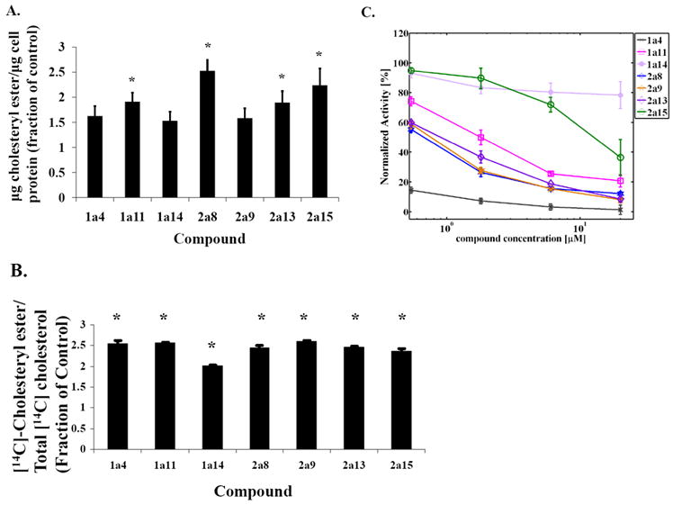
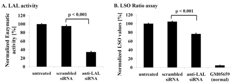
Niemann-Pick C disease (NPC) is a lysosomal storage disorder causing abnormal accumulation of unesterified free cholesterol in lysosomal storage organelles. High content phenotypic microscopy chemical screens in both human and hamster NPC-deficient cells have identified several compounds that partially revert the NPC phenotype. Cell biological and biochemical studies show that several of these molecules inhibit lysosomal acid lipase, the enzyme that hydrolyzes LDL-derived triacylglycerol and cholesteryl esters. The effects of reduced lysosomal acid lipase activity in lowering cholesterol accumulation in NPC mutant cells were verified by RNAi-mediated knockdown of lysosomal acid lipase in NPC1-deficient human fibroblasts. This work demonstrates the utility of phenotypic cellular screens as a means to identify molecular targets for altering a complex process such as intracellular cholesterol trafficking and metabolism.


Figures

Figure 1
Figure 1. Effect of compounds on NPC1 mutant and wild type CHO cells
(A) Quantification of cholesteryl ester levels by GC in CT60 cells treated with compounds. CT60 cells were treated with compounds (10 μM) overnight at 37 °C and cellular lipids were extracted. Total cholesterol (after saponification) and free cholesterol were measured, and the difference between these values was taken as the amount of cholesteryl ester. The content of cholesteryl ester in the cells per μg cell protein was determined for compound-treated cells and compared to the levels in control, DMSO-treated cells. (Referencing all values to the solvent-treated cells allowed us to pool data from separate experiments for statistical analysis.) For solvent-treated (control) CT60 cells, the mean value was 0.042 ± 0.004 μg cholesteryl ester/μg cell protein. For compound-treated cells, cholesteryl esters levels are presented as fractions of the control value and are shown as mean ± S.E. (* p < 0.01). Two to four independent experiments were conducted (6 ≤ n ≤ 12 per condition). (B) Hydrolysis of cholesteryl-[4-14C]-oleate-containing LDL in CT60 cells treated with compounds. CT60 cells were incubated overnight at 37 °C in 5 % LPDS medium and then pulsed for 2 h with 5 μ/ml cholesteryl-[14C]-oleate-LDL, compounds (10 μM) and 10 μg/ml ACAT inhibitor (compound 58-035). Afterward, cells were chased for 2 h at 37 °C in 5 % LPDS-medium with compounds (10 μM) and 10 μg/ml ACAT inhibitor. Lipids were extracted, and cholesteryl-[14C]-oleate was measured as a fraction of the total radiolabeled cholesterol. For DMSO-treated cells, the value was 0.37 ± 0.03. Experimental data of compound-treated cells are displayed as fractions of the control value and reflect averages ± S.E from three experiments (* p < 0.001, when compared to control; 6 ≤ n ≤ 9 per condition). (C) Lysosomal acid lipase activity in presence of compounds from the CHO cell screen. Triton X-100 cell extracts (~ 0.2 mg/ml) from TRVb1 cells were mixed with compounds at indicated final concentrations, and the reaction was started by adding 4MUO substrate. Enzymatic activity in the linear range of the assay at 37 °C was quantified as background corrected 4-methylumbelliferone fluorescence, normalized to DMSO control average value for a particular experiment. Data reflect averages ± S.E from two independent experiments (4 ≤ n ≤ 12 per condition).
Figure 2
Figure 2. siRNA knock-down of LAL
siRNA-mediated decrease in LAL expression as quantified by the 4MUO activity assay is illustrated in (A) and corresponding decrease in LSO values, as measured by the filipin staining and quantified by the LSO assay, is shown in (B). Data were obtained ~3 days post treatment with siRNA/Hiperfect complexes. LSO values were quantified using a specific threshold to select the LSOs and then normalized to the untreated control average value for a particular experiment. GM05659 cells are apparently normal fibroblasts. Enzymatic activity at 37 °C was quantified as background corrected increase in 4-methylumbelliferone fluorescence as a function of time (RFU/sec), normalized to protein concentration as measured by bicinchoninic acid assay (OD562), and subsequently normalized to the untreated control average value for a particular experiment. Data reflect averages ± S.E from five independent experiments (n ~ 40, per condition).
Figure 3
Figure 3. Lysosomal acid lipase activity in presence of compounds from the human NPC mutant cells screen
Compounds from GM03123 fibroblast screen (3a1, 3a2, 3a6, 3a7 and 3a8) show dose dependent inhibition of human purified enzyme, phLAL (A) and CHO (TRVb1) cell lysates (B). Human purified enzyme (phLAL, 0.003 U/ml, 105 U/mg) or Triton X-100 cell extracts (0.4–1.0 mg/ml) from TRVb1 cells was mixed with compounds at indicated final concentrations, and the reaction was started by adding 4MUO substrate. Enzymatic activity at 37 °C was quantified as background corrected 4-methylumbelliferone fluorescence, normalized to DMSO control average value for a particular experiment. Data reflect averages ± S.E from two independent experiments (n=28 for DMSO, n=6 for each compound (A); n=16 for DMSO, n=6 for each compound (B)).
Figure 4
Figure 4. Filipin images of GM03123 cells treated with LAL inhibitors show decreased cholesterol accumulation in LSOs
Cells were plated in 384-well plates in growth medium, and the next day treated with DMSO (A), 10 μM 3a2 (B), or 10 μM 3a7 (C) in screening medium (MEM, 5.5 % FBS, 10 mM HEPES, 2.2 g/L sodium bicarbonate, pH 7.4). After an overnight incubation, cells were washed with PBS, fixed with PFA and stained with filipin. 10X magnification images were obtained using ImageXpressMICRO automated imaging system. Images were corrected for shade and background. Bar = 100 μm. γ=0.74 was used to generate this figure.
Figure 5
Figure 5. LSO assay quantification of the effect of LAL inhibitors 3a1, 3a2, 3a6 and 3a7 on filipin-stained cholesterol in NPC mutant human fibroblasts (GM03123) and CHO (CT60) cells
GM03123 cells (A) or CT60 cells (B) were seeded in two 384-well plates in growth medium. After one day, compounds in screening medium were added to achieve final concentrations of 10 μM, 1 μM, 0.1 μM and 0.01 μM in 12 different wells per plate (total n=24/condition) and allowed to incubate overnight. Cells were washed with PBS, fixed with PFA and stained with filipin. 10X magnification filipin images were acquired and processed as described previously [38]. LSO ratio values reflecting cholesterol accumulation were calculated as described previously [38] and were normalized to control (DMSO-treated) average values. Data reflect averages ± S.E (when visible) from one representative experiment (n = 24 per compound condition).
Figure 6
Figure 6. Effect of LAL inhibition with compound 3a2 on the accumulation of neutral lipids in normal human fibroblasts
GM05659 cells were grown in growth medium with 10 % LPDS for 1 day, then treated with either 10 μM compound 3a2 for an additional day (A) or with DMSO (B), all in presence of 10 % LPDS and 100 μg/ml human LDL labeled with AlexaFluor-633 (red). Cells were then fixed with PFA and stained with LipidTOX (green). Confocal image stacks were acquired on a Zeiss LSM510 confocal microscope, and single optical sections of representative images are shown. For each channel, the same brightness and contrast settings were used to display images of compound-treated and control cells. Bar = 20 μm.
Figure 7
Figure 7. Time-course studies of compound pre-incubation with LAL
Compounds 3a1, 3a2, 3a6, and 3a7 were tested for time dependence in their inhibition of human purified enzyme, phLAL (0.003 U/ml, 105 U/mg). phLAL was incubated at room temperature for either ~ 2 hours or ~ 5 minutes with compounds at 50 nM final concentration, and the reaction was started by adding 4MUO substrate. Enzymatic activity at 37 °C was quantified as background corrected 4-methylumbelliferone fluorescence, normalized to DMSO control average value for a particular experiment. Data reflect averages ± S.E from two independent experiments, except for compound 3a1, where data from only one experiment are shown (3 ≤ n ≤ 17).
Figure 8
Figure 8. Human pancreatic lipase and bovine milk lipoprotein lipase activity assays in the presence of compounds selected from the human NPC mutant cells screen
Purified enzymes, human pancreatic lipase (2 U/ml, 468 U/mg) (A) and bovine milk lipoprotein lipase (465.3 U/ml, 5770 U/mg) (B), were mixed with 10 μM compounds, and the reaction was started by adding 4MUO substrate. Enzymatic activity at 37 °C was quantified as background corrected 4-methylumbelliferone fluorescence, normalized to the DMSO control average value for a particular experiment. Data reflect averages ± S.E from two independent experiments (5 ≤ n ≤ 12 per condition).

Similar articles

Cited by

References

    1. Patterson MC, Vanier MT, Suzuki K, Morris JA, Carstea E, Neufeld EB, Blanchette-Mackie EJ, Pentchev PG. Niemann-Pick Disease TypeC: A lipid Trafficking Disorder. In: Scriver CR, Beaudet AL, Sly WS, Valle D, editors. The Metabolic and Molecular Basis of Inherited Disease. Vol. 3. McGraw-Hill; New York: 2001. pp. 3611–3633.
    1. Maxfield FR, Tabas I. Role of cholesterol and lipid organization in disease. Nature. 2005;438:612–621. - PubMed
    1. Mukherjee S, Maxfield FR. Lipid and cholesterol trafficking in NPC. Biochim Biophys Acta. 2004;1685:28–37. - PubMed
    1. Pagano RE. Endocytic trafficking of glycosphingolipids in sphingolipid storage diseases. Philos Trans R Soc Lond B Biol Sci. 2003;358:885–891. - PMC - PubMed
    1. Sturley SL, Patterson MC, Balch W, Liscum L. The pathophysiology and mechanisms of NP-C disease. Biochim Biophys Acta. 2004;1685:83–87. - PubMed

Publication types

MeSH terms