Review
. 2009 Nov-Dec;54(6):686-96.
doi: 10.1016/j.survophthal.2009.03.004. Epub 2009 Aug 21.

Amniotic membrane transplantation as a new therapy for the acute ocular manifestations of Stevens-Johnson syndrome and toxic epidermal necrolysis

Affiliations
Review

Amniotic membrane transplantation as a new therapy for the acute ocular manifestations of Stevens-Johnson syndrome and toxic epidermal necrolysis

Elizabeth Shay et al. Surv Ophthalmol. 2009 Nov-Dec.

Abstract

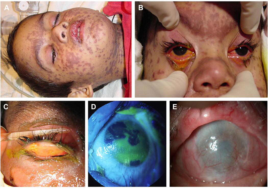
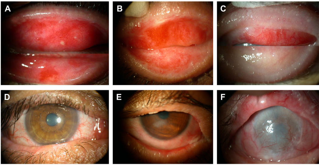
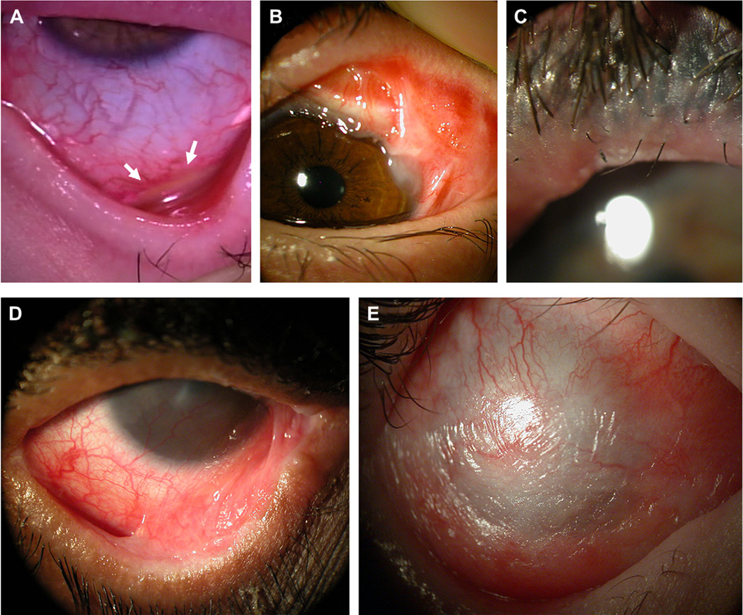
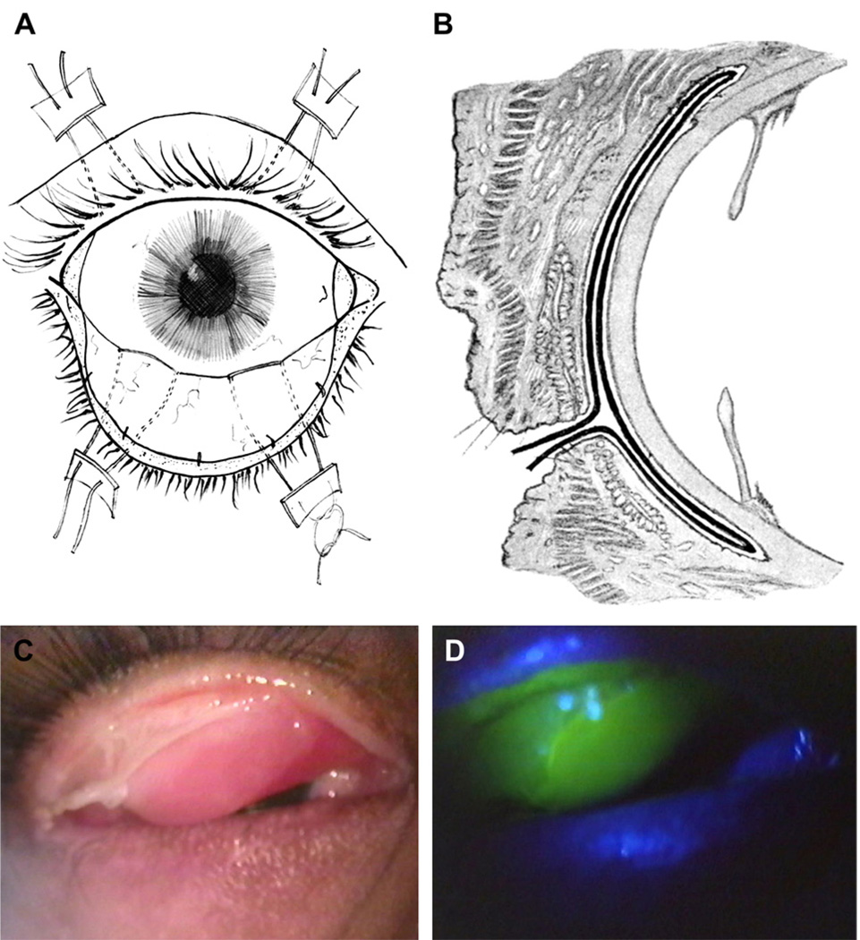
Stevens-Johnson syndrome and its more severe variant, toxic epidermal necrolysis, have relatively low overall incidence; however, this disease presents with high morbidity and mortality. The majority of patients develop ocular inflammation and ulceration at the acute stage. Due to the hidden nature of these ocular lesions and the concentration of effort toward life-threatening issues, current acute management has not devised a strategy to preclude blinding cicatricial complications. This review summarizes recent literature data, showing how sight-threatening corneal complications can progressively develop from cicatricial pathologies of lid margin, tarsus, and fornix at the chronic stage. It illustrates how such pathologies can be prevented with the early intervention of cryopreserved amniotic membrane transplantation to suppress inflammation and promote epithelial healing at the acute stage. Significant dry eye problems and photophobia can also be avoided with this intervention. This new therapeutic strategy can avert the catastrophic ophthalmic sequelae of this rare but devastating disease.


Figures

Fig. 1
Fig. 1
Acute ocular manifestation of SJS/TEN. A: External appearance of a SJS patient shows diffuse skin rashes, oral mucosal ulceration, and closure of both eyes with crust. B: External examination at the bedside reveals only ulcers involving the lid margin skin and conjunctival redness. (Reproduced from Di Pascuale et al with permission of Ophthalmology.) C: Eversion of the eyelids reveals diffuse ulcers involving the tarsus and fornix. D: Acute ocular involvement manifesting a geographical corneal epithelial defect. (C and D are reproduced from Kobayashi et al with permission of Ophthalmology.) E: Limbal stem cell deficiency can occur acutely, resulting in conjunctivalization and formation of an extensive fibrovascular scar, causing blindness.
Fig. 2
Fig. 2
Significant correlation between lid margin or tarsal keratinization and scarring with corneal scarring and vascularization. The severity of grade 1 (A), grade 2 (B), and grade 3 (C) of lid margin and tarsal scarring and keratinization of representative cases is correlated well with their corneas, which showed clear (D), mild scarring (E), and severe scarring and vascularization (F). (Reproduced from Di Pascuale et al with permission of Ophthalmology.)
Fig. 3
Fig. 3
Chronic cicatricial complications of SJS/TEN. A: Progressive conjunctival inflammation and non-healing fornix ulcer (marked by arrows) in the chronic stage, seen when the eyelid was everted by a muscle hook. B: Persistent conjunctival inflammation and scarring involves the superotemporal fornix, obstructing the lacrimal ductules. C: Lid margin keratinization and scarring leads to distichiasis and meibomian gland obstruction. D: Inferior symblepharon obliterates the tear meniscus, interfering with aqueous tear flow and spread to the ocular surface E: Diffuse squamous metaplasia due to severe dry eye and exposure.
Fig. 4
Fig. 4
Schematic depiction of the sutured method of AMT for SJS/TEN. AM covers the entire ocular surface, secured by four 4-0 silk double-armed sutures from the fornix to the skin (A: front view; B: side view.) (A and B are reproduced from Meller et al with permission of Ophthalmology.) C: AM is attached to the ulcerated tarsus while dissolving in other areas. D: Fluorescein staining confirms that AM has covered the tarsus. This membrane facilitates conjunctival epithelialization while preventing epidermal migration from the lid margin. (C and D are reproduced from Di Pascuale et al with permission of Ophthalmology.)

Similar articles

Cited by

References

    1. Adds PJ, Hunt CJ, Dart JK. Amniotic membrane grafts, “fresh” or frozen? A clinical and in vitro comparison. Br J Ophthalmol. 2001;85:905–907. - PMC - PubMed
    1. Aihara Y, Ito R, Ito S, et al. Toxic epidermal necrolysis in a child successfully treated with cyclosporin A and methyl-prednisolone. Pediatr Int. 2007 Oct;49(5):659–662. - PubMed
    1. Arevalo JM, Lorente JA, Gonzalez-Herrada C, Jimenez-Reyes J. Treatment of toxic epidermal necrolysis with cyclosporin A. J Trauma. 2000;48(3):473–478. - PubMed
    1. Arora R, Mehta D, Jain V. Amniotic membrane transplantation in acute chemical burns. Eye. 2005 Mar;19(3):273–278. - PubMed
    1. Arstikaitis MJ. Ocular aftermath of Stevens-Johnson syndrome. Arch Ophthalmol. 1973 Nov;90(5):376–379. - PubMed

Publication types