

Partially nanofibrous architecture of 3D tissue engineering scaffolds
- PMID: 19699518
- PMCID: PMC2763581
- DOI: 10.1016/j.biomaterials.2009.08.012
Partially nanofibrous architecture of 3D tissue engineering scaffolds
Abstract
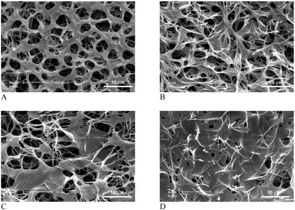
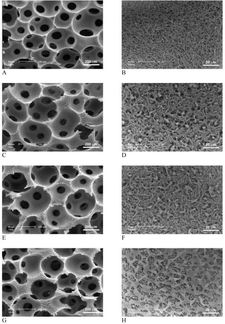
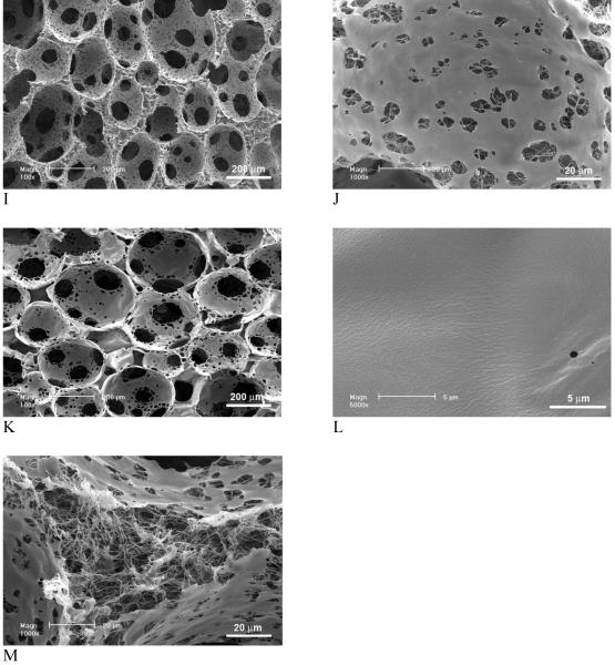
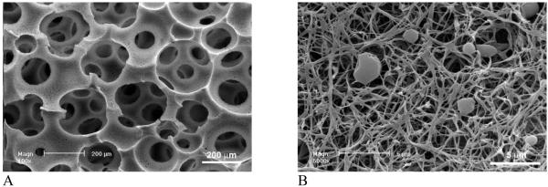
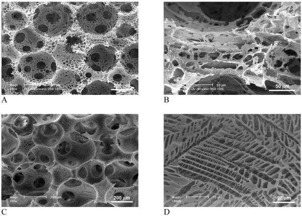
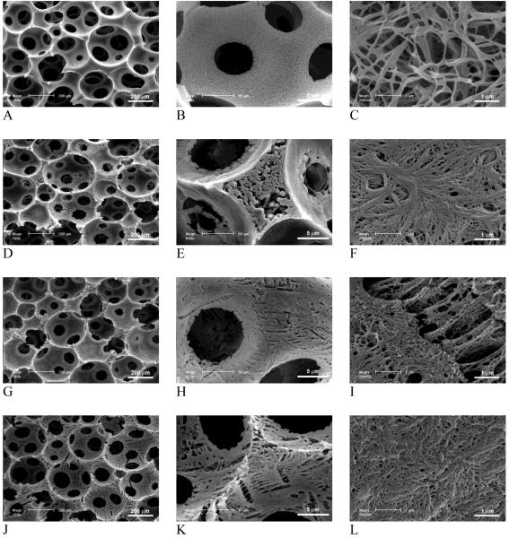
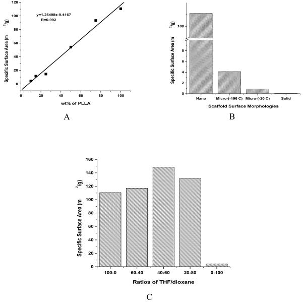
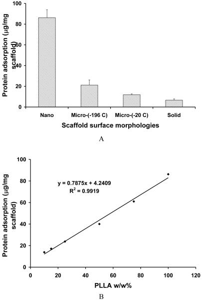
An ideal tissue-engineering scaffold should provide suitable pores and appropriate pore surface to induce desired cellular activities and to guide 3D tissue regeneration. In the present work, we have developed macroporous polymer scaffolds with varying pore wall architectures from smooth (solid), microporous, partially nanofibrous, to entirely nanofibrous ones. All scaffolds are designed to have well-controlled interconnected macropores, resulting from leaching sugar sphere template. We examine the effects of material composition, solvent, and phase separation temperature on the pore surface architecture of 3D scaffolds. In particular, phase separation of PLLA/PDLLA or PLLA/PLGA blends leads to partially nanofibrous scaffolds, in which PLLA forms nanofibers and PDLLA or PLGA forms the smooth (solid) surfaces on macropore walls, respectively. Specific surface areas are measured for scaffolds with similar macroporosity but different macropore wall architectures. It is found that the pore wall architecture predominates the total surface area of the scaffolds. The surface area of a partially nanofibrous scaffold increases linearly with the PLLA content in the polymer blend. The amounts of adsorbed proteins from serum increase with the surface area of the scaffolds. These macroporous scaffolds with adjustable pore wall surface architectures may provide a platform for investigating the cellular responses to pore surface architecture, and provide us with a powerful tool to develop superior scaffolds for various tissue-engineering applications.
Figures
Similar articles
-
Nano-fibrous poly(L-lactic acid) scaffolds with interconnected spherical macropores.Biomaterials. 2004 May;25(11):2065-73. doi: 10.1016/j.biomaterials.2003.08.058. Biomaterials. 2004. PMID: 14741621
-
Electrospun nanostructured scaffolds for bone tissue engineering.Acta Biomater. 2009 Oct;5(8):2884-93. doi: 10.1016/j.actbio.2009.05.007. Epub 2009 May 15. Acta Biomater. 2009. PMID: 19447211
-
Porogen-induced surface modification of nano-fibrous poly(L-lactic acid) scaffolds for tissue engineering.Biomaterials. 2006 Jul;27(21):3980-7. doi: 10.1016/j.biomaterials.2006.03.008. Epub 2006 Mar 31. Biomaterials. 2006. PMID: 16580063
-
Biomimetic and bioactive nanofibrous scaffolds from electrospun composite nanofibers.Int J Nanomedicine. 2007;2(4):623-38. Int J Nanomedicine. 2007. PMID: 18203429 Free PMC article. Review.
-
Nanostructured polymer scaffolds for tissue engineering and regenerative medicine.Wiley Interdiscip Rev Nanomed Nanobiotechnol. 2009 Mar-Apr;1(2):226-36. doi: 10.1002/wnan.26. Wiley Interdiscip Rev Nanomed Nanobiotechnol. 2009. PMID: 20049793 Free PMC article. Review.
Cited by
-
Delivery of PDGF-B and BMP-7 by mesoporous bioglass/silk fibrin scaffolds for the repair of osteoporotic defects.Biomaterials. 2012 Oct;33(28):6698-708. doi: 10.1016/j.biomaterials.2012.06.021. Epub 2012 Jul 2. Biomaterials. 2012. PMID: 22763224 Free PMC article.
-
Nanoscale Control of Silks for Nanofibrous Scaffold Formation with Improved Porous Structure.J Mater Chem B. 2014 May 7;2(17):2622-2633. doi: 10.1039/C4TB00019F. J Mater Chem B. 2014. PMID: 24949200 Free PMC article.
-
Urine-derived stem cells for potential use in bladder repair.Stem Cell Res Ther. 2014 May 28;5(3):69. doi: 10.1186/scrt458. Stem Cell Res Ther. 2014. PMID: 25157812 Free PMC article. Review.
-
Nanofiber-microsphere (nano-micro) matrices for bone regenerative engineering: a convergence approach toward matrix design.Regen Biomater. 2014 Nov;1(1):3-9. doi: 10.1093/rb/rbu002. Epub 2014 Oct 20. Regen Biomater. 2014. PMID: 26816620 Free PMC article.
-
Rapid Fabrication of Anatomically-Shaped Bone Scaffolds Using Indirect 3D Printing and Perfusion Techniques.Int J Mol Sci. 2020 Jan 2;21(1):315. doi: 10.3390/ijms21010315. Int J Mol Sci. 2020. PMID: 31906530 Free PMC article.
References
-
- Ma PX. Tissue Engineering. In: Kroschwitz JI, editor. Encyclopedia of Polymer Science and Technology. Third ed. John Wiley & Sons, Inc.; Hoboken, NJ: 2005. pp. 261–291.
-
- Ma PX. Scaffolds for tissue fabrication. Materials Today. 2004;7(5):30–40.
-
- Chen VJ, Ma PX. Nano-fibrous poly(L-lactic acid) scaffolds with interconnected spherical macropores. Biomaterials. 2004;25(11):2065–2073. - PubMed
-
- Ma PX, Zhang RY. Microtubular architecture of biodegradable polymer scaffolds. J Biomed Mater Res. 2001;56:469–477. - PubMed
-
- Muschler GF, Nakamoto C, Griffith LG. Engineering principles of clinical cell-based tissue engineering. J Bone Joint Surg Am. 2004;86-A(7):1541–1558. - PubMed
Publication types
MeSH terms
Substances
Grants and funding
LinkOut - more resources
Full Text Sources
