

Oxidized lipids enhance RANKL production by T lymphocytes: implications for lipid-induced bone loss
- PMID: 19699688
- PMCID: PMC2805272
- DOI: 10.1016/j.clim.2009.07.011
Oxidized lipids enhance RANKL production by T lymphocytes: implications for lipid-induced bone loss
Abstract
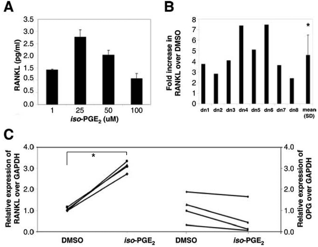
Osteoporosis is a systemic disease that is associated with increased morbidity, mortality and health care costs. Whereas osteoclasts and osteoblasts are the main regulators of bone homeostasis, recent studies underscore a key role for the immune system, particularly via activation-induced T lymphocyte production of receptor activator of NFkappaB ligand (RANKL). Well-documented as a mediator of T lymphocyte/dendritic cell interactions, RANKL also stimulates the maturation and activation of bone-resorbing osteoclasts. Given that lipid oxidation products mediate inflammatory and metabolic disorders such as osteoporosis and atherosclerosis, and since oxidized lipids affect several T lymphocyte functions, we hypothesized that RANKL production might also be subject to modulation by oxidized lipids. Here, we show that short term exposure of both unstimulated and activated human T lymphocytes to minimally oxidized low density lipoprotein (LDL), but not native LDL, significantly enhances RANKL production and promotes expression of the lectin-like oxidized LDL receptor-1 (LOX-1). The effect, which is also observed with 8-iso-Prostaglandin E2, an inflammatory isoprostane produced by lipid peroxidation, is mediated via the NFkappaB pathway, and involves increased RANKL mRNA expression. The link between oxidized lipids and T lymphocytes is further reinforced by analysis of hyperlipidemic mice, in which bone loss is associated with increased RANKL mRNA in T lymphocytes and elevated RANKL serum levels. Our results suggest a novel pathway by which T lymphocytes contribute to bone changes, namely, via oxidized lipid enhancement of RANKL production. These findings may help elucidate clinical associations between cardiovascular disease and decreased bone mass, and may also lead to new immune-based approaches to osteoporosis.
Figures

Similar articles
-
Oxidized low density lipoprotein enhanced RANKL expression in human osteoblast-like cells. Involvement of ERK, NFkappaB and NFAT.Biochim Biophys Acta. 2013 Oct;1832(10):1756-64. doi: 10.1016/j.bbadis.2013.05.033. Epub 2013 Jun 10. Biochim Biophys Acta. 2013. PMID: 23756197
-
Bone density and hyperlipidemia: the T-lymphocyte connection.J Bone Miner Res. 2010 Nov;25(11):2460-9. doi: 10.1002/jbmr.148. J Bone Miner Res. 2010. PMID: 20533376 Free PMC article.
-
Lectin-like oxidized low-density lipoprotein receptor-1 abrogation causes resistance to inflammatory bone destruction in mice, despite promoting osteoclastogenesis in the steady state.Bone. 2015 Jun;75:170-82. doi: 10.1016/j.bone.2015.02.025. Epub 2015 Mar 2. Bone. 2015. PMID: 25744064
-
[The OPG/RANKL/RANK system and bone resorptive disease].Sheng Wu Gong Cheng Xue Bao. 2003 Nov;19(6):655-60. Sheng Wu Gong Cheng Xue Bao. 2003. PMID: 15971575 Review. Chinese.
-
Immune response: the key to bone resorption in periodontal disease.J Periodontol. 2005 Nov;76(11 Suppl):2033-41. doi: 10.1902/jop.2005.76.11-S.2033. J Periodontol. 2005. PMID: 16277573 Review.
Cited by
-
Functional impairment of bone formation in the pathogenesis of osteoporosis: the bone marrow regenerative competence.Curr Osteoporos Rep. 2013 Jun;11(2):117-25. doi: 10.1007/s11914-013-0139-2. Curr Osteoporos Rep. 2013. PMID: 23471774 Review.
-
Neutralization of oxidized phospholipids attenuates age-associated bone loss in mice.Aging Cell. 2021 Aug;20(8):e13442. doi: 10.1111/acel.13442. Epub 2021 Jul 19. Aging Cell. 2021. PMID: 34278710 Free PMC article.
-
Association of Decreased Bone Density and Hyperlipidemia in a Taiwanese Older Adult Population.J Endocr Soc. 2024 Mar 5;8(5):bvae035. doi: 10.1210/jendso/bvae035. eCollection 2024 Mar 12. J Endocr Soc. 2024. PMID: 38505562 Free PMC article.
-
A Neutralizing Antibody Targeting Oxidized Phospholipids Promotes Bone Anabolism in Chow-Fed Young Adult Mice.J Bone Miner Res. 2021 Jan;36(1):170-185. doi: 10.1002/jbmr.4173. Epub 2020 Sep 29. J Bone Miner Res. 2021. PMID: 32990984 Free PMC article.
-
Obesity and lipid metabolism in the development of osteoporosis (Review).Int J Mol Med. 2024 Jul;54(1):61. doi: 10.3892/ijmm.2024.5385. Epub 2024 May 31. Int J Mol Med. 2024. PMID: 38818830 Free PMC article. Review.
References
-
- Burge R, Dawson-Hughes B, Solomon DH, Wong JB, King A, Tosteson A. Incidence and economic burden of osteoporosis-related fractures in the United States, 2005–2025. J Bone Miner Res. 2007;22:465–475. - PubMed
-
- Kanis JA. Diagnosis of osteoporosis and assessment of fracture risk. Lancet. 2002;359:1929–1936. - PubMed
-
- Walsh MC, Kim N, Kadono Y, Rho J, Lee SY, Lorenzo J, Choi Y. Osteoimmunology: interplay between the immune system and bone metabolism. Annu Rev Immunol. 2006;24:33–63. - PubMed
-
- Arron JR, Choi Y. Bone versus immune system. Nature. 2000;408:535–536. - PubMed
-
- Wong BR, Rho J, Arron J, Robinson E, Orlinick J, Chao M, Kalachikov S, Cayani E, Bartlett FS, 3rd, Frankel WN, Lee SY, Choi Y. TRANCE is a novel ligand of the tumor necrosis factor receptor family that activates c-Jun N-terminal kinase in T cells. J Biol Chem. 1997;272:25190–25194. - PubMed
Publication types
MeSH terms
Substances
Grants and funding
LinkOut - more resources
Full Text Sources
