. 2009 Oct 2;8(10):1235-41.
doi: 10.1016/j.dnarep.2009.07.007. Epub 2009 Aug 21.

Regulation of repair choice: Cdk1 suppresses recruitment of end joining factors at DNA breaks

Affiliations

Regulation of repair choice: Cdk1 suppresses recruitment of end joining factors at DNA breaks

Yu Zhang et al. DNA Repair (Amst). .

Abstract

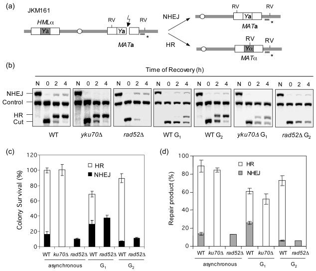
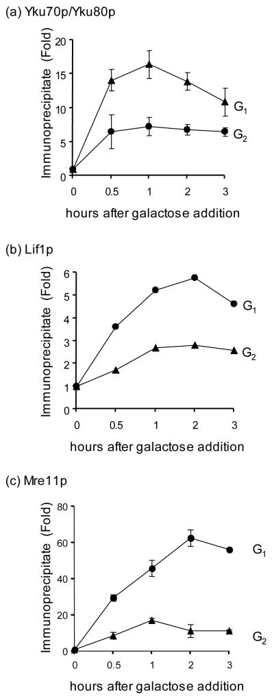
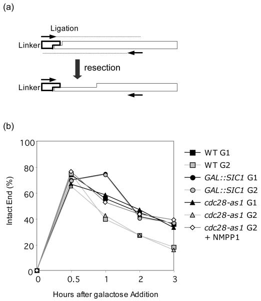
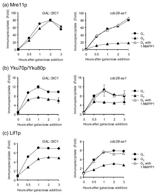
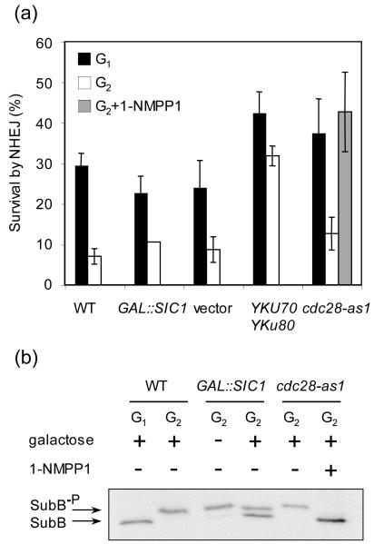
Cell cycle plays a crucial role in regulating the pathway used to repair DNA double-strand breaks (DSBs). In Saccharomyces cerevisiae, homologous recombination is primarily limited to non-G(1) cells as the formation of recombinogenic single-stranded DNA requires CDK1-dependent 5' to 3' resection of DNA ends. However, the effect of cell cycle on non-homologous end joining (NHEJ) is not yet clearly defined. Using an assay to quantitatively measure the contributions of each repair pathway to repair product formation and cellular survival after DSB induction, we found that NHEJ is most efficient at G(1), and markedly repressed at G(2). Repression of NHEJ at G(2) is achieved by efficient end resection and by the reduced association of core NHEJ proteins with DNA breaks, both of which depend on the CDK1 activity. Importantly, repression of 5' end resection by CDK1 inhibition at G(2) alone did not fully restore either physical association of Ku/Dnl4-Lif1 with DSBs or NHEJ proficiency to the level at G(1). Expression of excess Ku can partially offset the inhibition of end joining at G(2). The results suggest that regulation of Ku/Dnl4-Lif1 affinity for DNA ends may contribute to the cell cycle-dependent modulation of NHEJ efficiency.


Figures

Figure 1
Figure 1
NHEJ is repressed at G2. (a) Schematic diagram showing relevant genomic structure of JKM161. The location of a MAT specific probe (*) and the restriction endonuclease cleavage sites (EcoRV: RV) used for Southern blot analysis to detect repair product formation are indicated. (b) Southern blot analysis of the repair product formation. N represents no galactose. (c) Plot demonstrating percent colony survival by HR and NHEJ. (d) Plot demonstrating percentage of repair products by HR and NHEJ. Percent repair product was calculated by dividing the repair product signal (HR or NHEJ in (b)) with the signal of the HIS3 control (control) after 4 h of recovery in the glucose containing medium. Data represents the mean ± s.d. of three or more independent experiments.
Figure 2
Figure 2
The recruitment of NHEJ proteins at DSBs is reduced at G2. Kinetics of Yku (a), Lif1 (b), and Mre11 (c) recruitment to the HO induced DSB in G1 and G2 were determined by ChIP assays. Fold immunoprecipitate represents the ratio of the Yku, Lif1, or Mre11 IP PCR signal before and after HO induction, normalized by the PCR signal of the PRE1 control. Data represent the mean ± s.d. of three or more independent experiments.
Figure 3
Figure 3
Cdk1 activity suppresses end resection. (a) Scheme of the ligation-mediated PCR (LM-PCR). The adaptor, fully complementary to the 4 nucleotide 3′ overhang generated by HO induced cleavage, is ligated to genomic DNA. The two primers (arrows) anneal to the adaptor and 0.4 kb distal to HO cut site and will only amplify DNA ends with intact, unresected 5′ chromosomal ends. Even a single base pair of resection will prevent the ligation and leave no signal in the PCR reaction. (b) Percentage of intact DNA ends remaining at HO cleavage site at the indicated time after HO induction. Genomic DNA isolated at each time interval after HO expression was subjected to LM-PCR as described in the Materials and Methods. Percent intact ends is shown as the ratio of the PCR signal from each ligation reaction sample before and after HO induction, normalized by the PCR signal of the PRE1 control. To calculate percent intact ends, the ratio of the LM-PCR signal from Lev473 strain carrying HO recognition sequences modified to a BstXI site, whose digestion generates the same 4 nucleotide 3′ overhang as HO cleavage, normalized by the PCR signal of the PRE1 control after BstXI cleavage, was set to 100% [25].
Figure 4
Figure 4
The recruitment of NHEJ proteins at DSBs is suppressed by the Cdk1 activity but not by end resection. Kinetics of Mre11 (a), Yku (b), and Lif1 (c) recruitment to the HO induced DSB in G1 and G2 from strains expressing galactose inducible Sic1 (left panel) or harboring cdc28-as1 allele supplemented with 1-NMPP1 inhibitor (right panel) were determined by ChIP assays. Fold immunoprecipitate was calculated as described in the legend for Fig. 2. Data represent the mean ± s.d. of two or more independent experiments.
Figure 5
Figure 5
NHEJ is repressed by Cdk1 activity. (a) Effect of Cdk1 inactivation on colony survival by NHEJ of an HO-induced DNA break. Cultures were induced to express HO for 1 h and plated onto YEPD medium, which shuts off HO expression. Percent survival by NHEJ is shown as the number of a type colonies growing onto the YEPD plates after 1 h of HO induction, normalized by the number of colonies growing before HO induction. Data represent the mean ± s.d. of three or more independent experiments. (b) Cdk1 activity was measured by the phosphorylation of the B subunit of DNA polymerase-alpha, a marker for Cdc28/Clb activity [11].

Similar articles

Cited by

References

    1. Kolodner RD, Putnam CD, Myung K. Maintenance of genome stability in Saccharomyces cerevisiae. Science. 2002;297:552–557. - PubMed
    1. Wyman C, Kanaar R. DNA double-strand break repair: all’s well that ends well. Annu Rev Genet. 2006;40:363–383. - PubMed
    1. Shrivastav M, De Haro LP, Nickoloff JA. Regulation of DNA double-strand break repair pathway choice. Cell Res. 2008;18:134–147. - PubMed
    1. Daley JM, Palmbos PL, Wu D, Wilson TE. Nonhomologous end joining in yeast. Annu Rev Genet. 2005;39:431–451. - PubMed
    1. Lieber MR, Ma Y, Pannicke U, Schwarz K. The mechanism of vertebrate nonhomologous DNA end joining and its role in V(D)J recombination. DNA Repair (Amst) 2004;3:817–826. - PubMed

Publication types

MeSH terms