. 2009 Nov 6;389(1):57-62.
doi: 10.1016/j.bbrc.2009.08.089. Epub 2009 Aug 21.

A novel fibronectin type III module binding motif identified on C-terminus of Leptospira immunoglobulin-like protein, LigB

Affiliations

A novel fibronectin type III module binding motif identified on C-terminus of Leptospira immunoglobulin-like protein, LigB

Yi-Pin Lin et al. Biochem Biophys Res Commun. .

Abstract

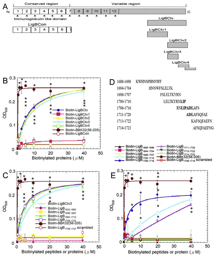
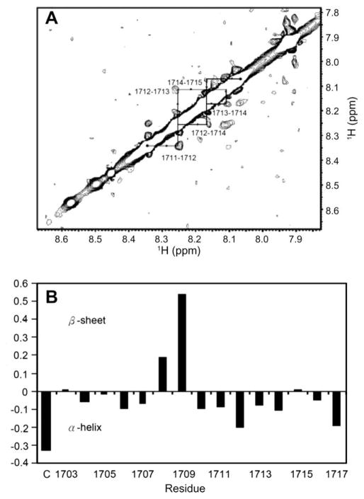
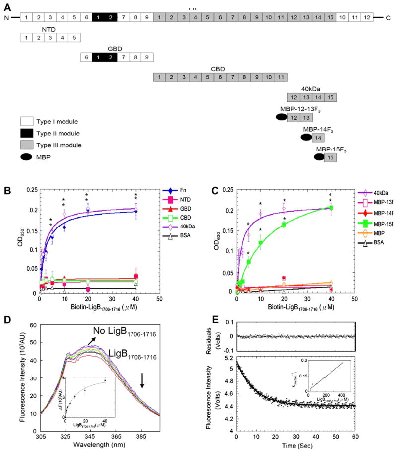
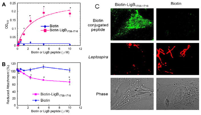
Infection by pathogenic strains of Leptospira hinges on the pathogen's ability to adhere to host cells via extracellular matrix such as fibronectin (Fn). Previously, the immunoglobulin-like domains of Leptospira Lig proteins were recognized as adhesins binding to N-terminal domain (NTD) and gelatin binding domain (GBD) of Fn. In this study, we identified another Fn-binding motif on the C-terminus of the Leptospira adhesin LigB (LigBCtv), residues 1708-1712 containing sequence LIPAD with a beta-strand and nascent helical structure. This motif binds to 15th type III modules (15F(3)) (K(D)=10.70 microM), and association (k(on)=600 M(-1)s(-1)) and dissociation (k(off)=0.0129 s(-1)) rate constants represents a slow binding kinetics in this interaction. Moreover, pretreatment of MDCK cells with LigB(1706-1716) blocked the binding of Leptospira by 39%, demonstrating a significant role of LigB(1706-1716) in cellular adhesion. These data indicate that the LIPAD residues (LigB(1708-1712)) of the Leptospira interrogans LigB protein bind 15F(3) of Fn at a novel binding site, and this interaction contributes to adhesion to host cells.


Figures

Fig. 1
Fig. 1
Identification of Fn-binding residues on LigBCtv. (A) A schematic diagram showing the truncated LigBCtv used in this study. (B) The Fn-binding activity of LigBCtv1, LigBCtv2, LigBCtv3, and LigBCtv4 regions. (C) The Fn-binding activity of the 30mers peptides from LigBCtv. (D) The synthetic peptide sequences (11mers) with overlapping regions used to identify the essential amino acids (indicated in bold) required for Fn binding. (E) The synthetic peptides indicated in (D) used in the Fn-binding assays. Various concentrations (40, 20, 10, 5, 2.5, 1.25, and 0.625 μM) of each tested biotinylated-protein or -peptide, -BBK3256–205 (positive control; B, C, and E), -LigBCon (negative control; B) or -scrambled peptide (negative control; C and E) were used in this study. Bound proteins or peptides were measured by ELISA.
Fig. 2
Fig. 2
(A) HN–HN region of the homonuclear NOESY spectrum (tmix = 200 ms, pH 6.0) of LigB1703–1717 in PBS in H2O. The dNN cross-peaks are labeled with residue numbers, and arrows indicate sequential connectivities. The NOEs between sequential residues in residues A1711 through F1715 indicate nascent helical character. (B) Plot of the difference in Hα chemical shifts, or Δδ, between observed and random coil values for LigB1703–1717. (C) Indicated the first cysteine conjugated on LigB1703–1717. A stretch of Δδ values below −0.1 indicates nascent α-helical structure, and a stretch of values above 0.1 indicates preference for β-strand conformation.
Fig. 3
Fig. 3
Mapping and characterization of LigB1706–1716 binding sites on Fn. (A) A chart presenting Fn and truncated Fn used in this study. (B,C) Various concentration (0.625, 1.25, 2.5, 5, 10, 20, 40 μM) of biotin-LigB1706–1716 or biotin (negative control and data not shown) were added to 1 μg of (B) Fn, NTD, GBD, CBD, 40 kDa, or BSA (negative control) (C) 40 kDa, MBP-12–13F3, MBP-14F3, MBP-15F3, MBP (negative control), or BSA (negative control) coated microtiter plate wells. Bound proteins were measured by ELISA. (D) 1 μM of 15F3 in PBS in the presence of 0, 0.625, 1.25, 2.5, 5, 10, 20 and 40 μM of LigB1706–1716 was excited at 295 nm to measure Trp fluorescence. Inner plot: KD of 15F3-LigB1706–1716 determined by monitoring quenching fluorescence intensities at 350 nm. (E) Stop flow experiment of 15F3 binding to LigB1706–1716. The signal represents the total fluorescence emission above 320 nm by excited at 295 nm. Inner plot: The kinetic plot of kobs versus concentration of 1 μM 15F3 under different LigB1706–1716 concentrations (25, 50, 100, 200, 400 μM).
Fig. 4
Fig. 4
The binding of LigB1706–1716 to MDCK cells reduces leptospiral adhesion. (A) Various concentrations (0, 2, 4, 6, 8, or 10 μM) of biotin-LigB1706–1716, or biotin (negative control) were added to MDCK cells (105). (B,C) LigB1706–1716 inhibits the binding of Leptospira to MDCK cells. (B) MDCK cells (105) were incubated with various concentrations (0, 2, 4, 6, or 8, 10 μM) of biotin-LigB1706–1716, or biotin (negative control) prior to the addition of Leptospira (107). (A) The binding or (B) the reduced attachment was measured by ELISA. (C) The adhesion of Leptospira (108) or the binding of 10 μM of biotin or biotin-LigB1706–1716 to MDCK cells (106) was detected by CLSM.

Similar articles

Cited by

References

    1. Faine SB, Adher B, Bolin C, et al. Leptospira and Leptospirosis. MedSci; Melbourne, Australia: 1999.
    1. Meites E, Jay MT, Deresinski S, et al. Reemerging leptospirosis, California. Emerg Infect Dis. 2004;10:406–412. - PMC - PubMed
    1. Schwarz-Linek U, Hook M, Potts JR. The molecular basis of fibronectin-mediated bacterial adherence to host cells. Mol Microbiol. 2004;52:631–641. - PubMed
    1. Lin YP, Chang YF. A domain of the Leptospira LigB contributes to high affinity binding of fibronectin. Biochem Biophys Res Commun. 2007;362:443–448. - PubMed
    1. Lin YP, Chang YF. The C-terminal variable domain of LigB from Leptospira mediates binding to fibronectin. J Vet Sci. 2008;9:133–144. - PMC - PubMed

Publication types

Substances