. 2009 Oct 15;334(2):481-90.
doi: 10.1016/j.ydbio.2009.08.015. Epub 2009 Aug 21.

Tgfbr2 is required for development of the skull vault

Affiliations

Tgfbr2 is required for development of the skull vault

Hwa-Seon Seo et al. Dev Biol. .

Abstract

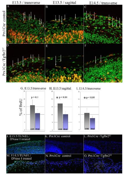
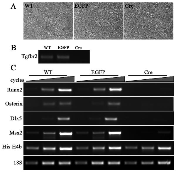
Transforming growth factor beta (TGFbeta) is known to play important roles in multiple developmental processes. One of the main functions is in skeletal development. Our previous studies demonstrated that loss of Tgfbr2 in Prx1Cre-expressing limb mesenchyme results in defects in the long bones and joints of mice. Here we show that loss of Tgfbr2 also results in defects in the development of the skull vault indicating Tgfbr2 has a critical role in intramembranous bone formation as well as endochondral bone formation. Mutant mice did not survive after birth and demonstrated an open skull. The first signs of skull defects were observed at E14.5 day. Prx1Cre(+)/Tgfbr2(f/f) embryos showed significantly reduced cell proliferation in the developing mesenchyme of the skull by E14.5 day without any detectable alteration in apoptosis suggesting that reduced cell proliferation in Prx1Cre(+)/Tgfbr2(f/f) embryos was at least partially responsible for the defects observed. Immunofluorescent staining showed a significant reduction in the expression of Runx2/Cbfa1 and Osterix/Sp7 in Prx1Cre(+)/Tgfbr2(f/f) embryos suggesting that osteoblast differentiation was also altered in Prx1Cre(+)/Tgfbr2(f/f) embryos. To distinguish between the effects of losing Tgfbr2 on mesenchymal proliferation versus osteoblast differentiation, osteoprogenitor cells from the skulls of Tgfbr2(f/f) embryos were cultured under conditions of high cell density and Tgfbr2 was deleted from the cells using Adeno-Cre virus. RT-PCR analysis showed that the mRNA level of Runx2 and Osterix as well as Dlx5 and Msx2 were down-regulated in Tgfbr2-deleted cultures compared to control cultures indicating that Tgfbr2 regulates osteoblast differentiation independent of regulating proliferation. Together, these results suggest that Tgfbr2 is required for normal development of the skull.


Figures

Figure 1
Figure 1. Tissue specific deletion of Tgfbr2
(A) To analyze deletion of Tgfbr2, mRNA was isolated from the developing skull mesenchyme of E14.5 Prx1Cre/ Tgfbr2f/w (−; f/w), and Prx1Cre+/ Tgfbr2f/f (+; f/f) embryos. Relative levels of Tgfbr2 mRNA were determined using RT-PCR. Primers recognizing sequences within exon2 were used. The level of Tgfbr2 mRNA was significantly reduced in Prx1Cre+/ Tgfbr2f/f embryos compared to Cre control embryos. Product formation is shown within the linear range. Expression of β2-microglobulin was used as a normalization control. (B-G) Prx1Cre recombinase activity was examined by X-gal staining in E13.5 (B-D) and 15.5 day embryos (E-G). Sagittal sections from E13.5 day Prx1Cre+/R26R embryos show strong Cre activity in the developing skull. In E15.5 day embryos, Cre activity was detected within differentiating osteoprogenitor cells (o) and in the dermis (d) in both frontal (F) and parietal (G) bones. Activity was not detected in the epidermis of the skin (s) or meninges (m) suggesting tissue specific deletion of Tgfbr2 in Prx1Cre+/Tgfbr2f/f embryos at these stages. fr= presumptive frontal bone, pa = presumptive parietal bone, br = brain
Figure 2
Figure 2. Skull defects in Prx1Cre+/Tgfbr2f/f embryo
(A) Prx1Cre+/Tgfbr2f/f (left) mice were born without a skull vault. Prx1Cre- control is on the right. (B-G) Alcian blue and Alizarin red staining shows details of skull defects in Prx1Cre+/Tgfbr2f/f (C, E, G) and Prx1Cre negative control embryos (B, D, F) at E15.5 days (B, C) and E18.5 days (D-G). At E15.5, control embryos (B) show distinctive frontal, parietal bone and sutural area which are not detected in Prx1Cre+/Tgfbr2f/f embryos (C). At E18.5, skeletal staining of control embryos show well developed frontal and parietal bone as well as base of skull (D, F). In Prx1Cre+/Tgfbr2f/f embryos, most of frontal and parietal bone are absent, but base of skull shows normal development compared to control embryos (E, G). (fr, frontal bone; pa, parietal bone; su, suture; ip, interparietal bone; na, nasal; oc, occipital bone; mx, maxilla; ma, mandible; zy, zygomatic; sq, squamosal)
Figure 3
Figure 3. Histological analysis of skull defects in Prx1Cre+/Tgfbr2f/f embryos
(A) Black and white lines indicate the sectioned area of skull. Sagital sections show the medial aspect and transverse sections show the laterals axpects of the skull. Transverse (B-K) and sagittal (L-O) sections of developing skull from Prx1Cre+/Tgfbr2f/f (C, E, G, I , K, M, O) and control embryos (B, D, F, H, J, L, N) were stained with H&E for histological analysis (A-G, L-O) or von Kossa to visualize mineral (H-K). At E13.5 day, the lateral aspect of the presumptive parietal bone in Prx1Cre+/Tgfbr2f/f (B, brackets) and control embryos (C, brackets) demonstrate similar thickness. E15.5 day Prx1Cre+/Tgfbr2f/f embryos showed a significant reduction in thickness (fr, pa, brackets) and mineralization (arrowheads) in frontal and parietal bone compared to control embryos (D-K). At E16.5 day, Prx1Cre+/Tgfbr2f/f embryos (M,O, arrowhead) demonstrated reduced thickness in presumptive frontal and parietal bones compared to control embryos (L, N, arrowhead). (fr, frontal bone; pa, parietal bone; m, meninges; ep, epidermis; d, dermis)
Figure 4
Figure 4. Reduced cell proliferation in presumptive skull bones
Immunostaining of BrdU-labeled osteoprogenitor cells in the developing parietal bone from E13.5 (A, B, D, E) and E14.5 (C, F) day control (A-C) and mutant (D-F) embryos. Specimens were immunostained using an anti-BrdU antibody (orange, arrows) and counterstained with YoPro (green). BrdU positive cells in skin and the brain were not counted. In the lateral aspects of the skull from E13.5 day embryos, percentages of proliferating cells were reduced but not statistically significant (A, D, G). Significant reduction in the number of BrdU positive cells was detected in the medial aspect of E13.5 day (B, E, H) and lateral aspect of E14.5 day (C, F, I) mutant embryos compared to control embryos. At E13.5 (J-L) and E14.5 (M-O) day, no apparent apoptotic activity was detected in either Prx1Cre+/Tgfbr2f/f (L,O) or control (K, N) embryos indicating that the failure of skull development in Prx1Cre+/Tgfbr2f/f embryos was not due to an increase in apoptosis but a decrease in cell proliferation. TUNEL positive signals are shown as green cells in the DNase I-treated positive control (J, M).
Figure 5
Figure 5. Expression of osteoblast markers was altered in Prx1Cre+/Tgfbr2f/f embryos
Expression of Runx2 and Osterix were examined in the presumptive parietal bone in E13.5 (A, B, G, H), E14.5 (C, D, I, J) and E15.5 (E, F, K, L) day embryos. Specimens were immunostained using an anti-Runx2 (A-F) and anti-Osterix antibodies (G-H) (orange, arrow) and counterstained with YoPro (green). At E13.5 and E14.5 day, control embryos show expression of Runx2 and Osterix in lateral aspects of the skull (A, C, G, I, arrow). In E13.5 day Prx1Cre+/Tgfbr2f/f embryos (B, H), Runx2 and Osterix expression in lateral aspects of skull was comparable with control embryos (A, G), but a significant reduction of Runx2 and Osterix expression was detected in E14.5 day Prx1Cre+/Tgfbr2f/f embryos compared to control embryos (C, D, I, J, arrow). In E15.5 day Prx1Cre+/Tgfbr2f/f embryos, there was no Runx2 and Osterix expression in the roof of the skull compared to control embryos (E, F, K, L, arrow) indicating altered osteoblast differentiation in Prx1Cre+/Tgfbr2f/f embryos.
Figure 6
Figure 6. Tgfbr2 regulates differentiation of osteoblasts independent of effects on proliferation
Mesenchymal cells from E14.5 day Tgfbr2f/f skulls were cultured to confluence then were uninfected (con) or infected with Ad-EGFP (EGFP) or Ad-Cre (Cre) adenovirus. Three days after infection there was no significant or consistent differences in the morphology of the cells (A). RT-PCR analysis showed effective deletion of Tgfbr2 in Ad-Cre virus treated culture (B). mRNA was also isolated from cultured cells for semi-quantitative RT-PCR analysis of osteoblast markers in cells with deletion of Tgfbr2 under conditions of high cell density (C). cDNA template was amplified for varying cycles number to ensure product formation in the linear range. Results are shown for product formation with varying cycle number (triangle illustrates increasing cycle number). The levels of Runx2/Cbfa1, Osterix/Sp7, Dlx5, Msx2 were significantly down regulated in Ad-Cre infected cells compared to Ad-EGFP infected and uninfected control cells (C). Histone H4b expression was similar in Ad-Cre and Ad-EGFP infected cells as well as in uninfected cells (C).

Similar articles

Cited by

References

    1. Acampora D, Merlo GR, Paleari L, Zerega B, Postiglione MP, Mantero S, Bober E, Barbieri O, Simeone A, Levi G. Craniofacial, vestibular and bone defects in mice lacking the Distal-less-related gene Dlx5. Development. 1999;126:3795–809. - PubMed
    1. Baffi MO, Slattery E, Sohn P, Moses HL, Chytil A, Serra R. Conditional deletion of the TGF-beta type II receptor in Col2a expressing cells results in defects in the axial skeleton without alterations in chondrocyte differentiation or embryonic development of long bones. Dev Biol. 2004;276:124–42. - PubMed
    1. Celil AB, Hollinger JO, Campbell PG. Osx transcriptional regulation is mediated by additional pathways to BMP2/Smad signaling. J Cell Biochem. 2005;95:518–28. - PubMed
    1. Chai Y, Jiang X, Ito Y, Bringas P, Jr., Han J, Rowitch DH, Soriano P, McMahon AP, Sucov HM. Fate of the mammalian cranial neural crest during tooth and mandibular morphogenesis. Development. 2000;127:1671–9. - PubMed
    1. Chytil A, Magnuson MA, Wright CV, Moses HL. Conditional inactivation of the TGF-beta type II receptor using Cre:Lox. Genesis. 2002;32:73–5. - PubMed

Publication types

MeSH terms

Substances

LinkOut - more resources