. 2009 Nov 1;335(1):93-105.
doi: 10.1016/j.ydbio.2009.08.014. Epub 2009 Aug 21.

BCL9 is an essential component of canonical Wnt signaling that mediates the differentiation of myogenic progenitors during muscle regeneration

Affiliations

BCL9 is an essential component of canonical Wnt signaling that mediates the differentiation of myogenic progenitors during muscle regeneration

Andrew S Brack et al. Dev Biol. .

Abstract

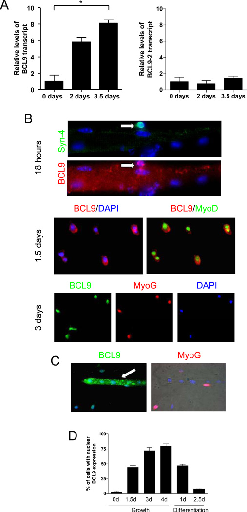
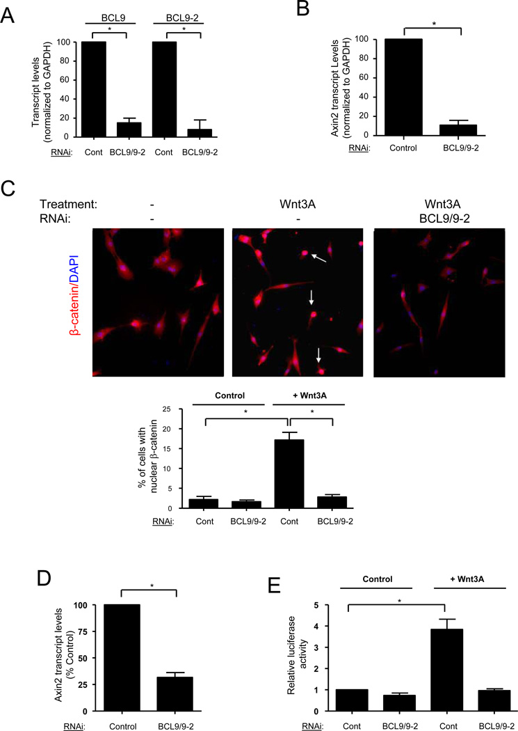
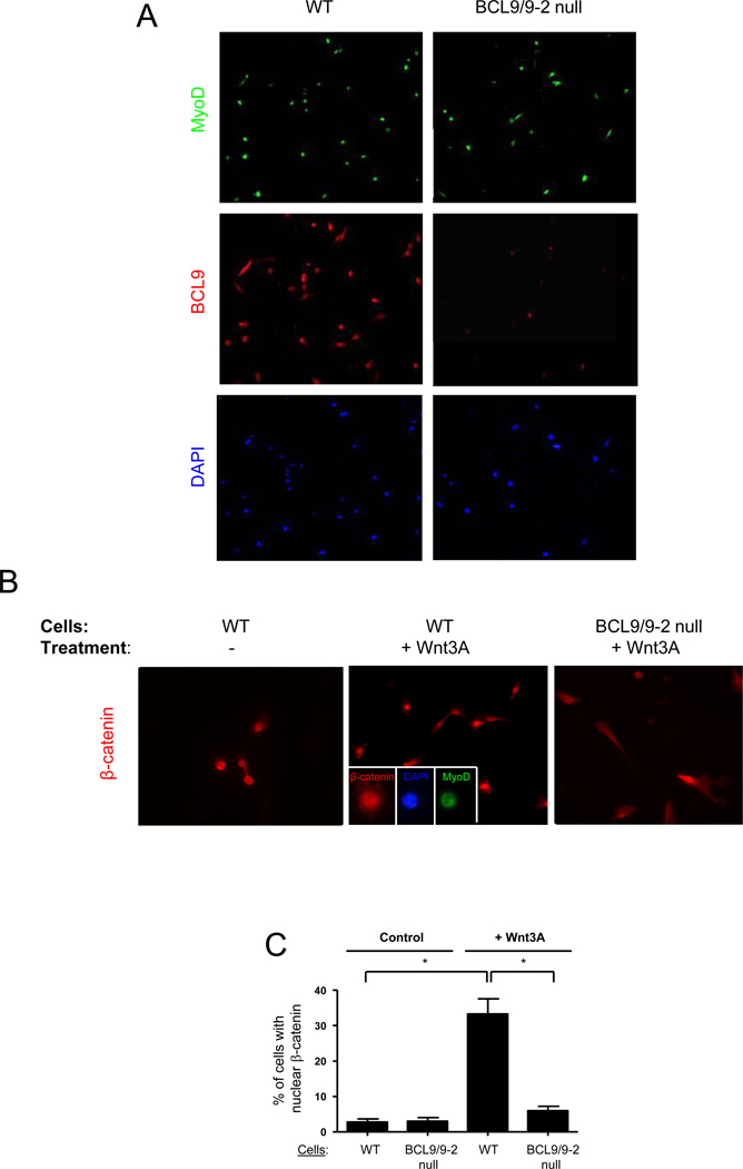
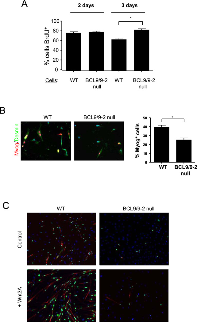
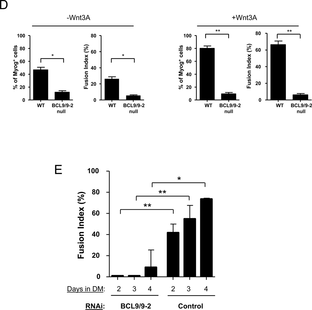
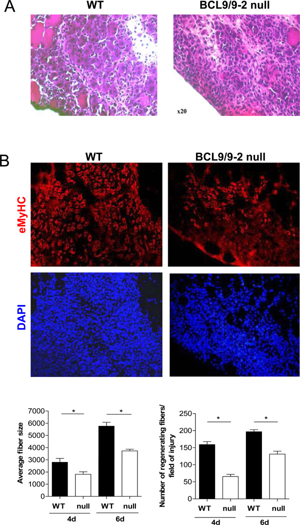
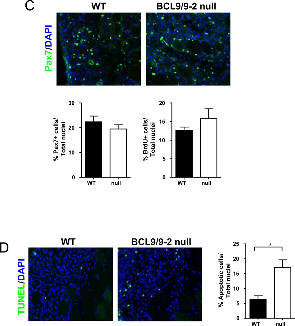
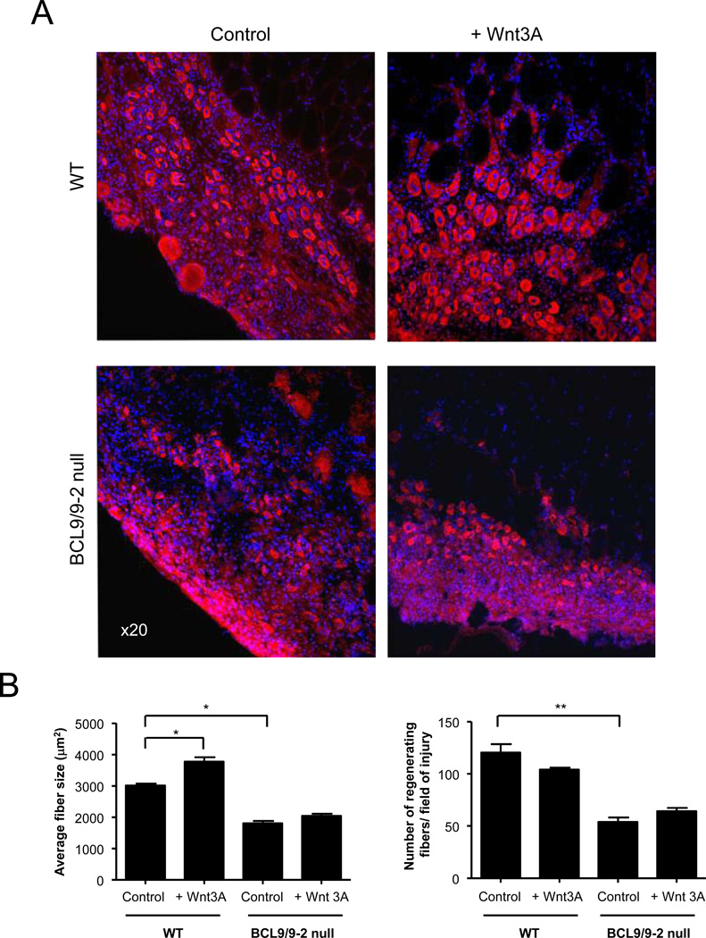
Muscle stem cells and their progeny play a fundamental role in the regeneration of adult skeletal muscle. We have previously shown that activation of the canonical Wnt/beta-catenin signaling pathway in adult myogenic progenitors is required for their transition from rapidly dividing transient amplifying cells to more differentiated progenitors. Whereas Wnt signaling in Drosophila is dependent on the presence of the co-regulator Legless, previous studies of the mammalian ortholog of Legless, BCL9 (and its homolog, BCL9-2), have not revealed an essential role of these proteins in Wnt signaling in specific tissues during development. Using Cre-lox technology to delete BCL9 and BCL9-2 in the myogenic lineage in vivo and RNAi technology to knockdown the protein levels in vitro, we show that BCL9 is required for activation of the Wnt/beta-catenin cascade in adult mammalian myogenic progenitors. We observed that the nuclear localization of beta-catenin and downstream TCF/LEF-mediated transcription, which are normally observed in myogenic progenitors upon addition of exogenous Wnt and during muscle regeneration, were abrogated when BCL9/9-2 levels were reduced. Furthermore, reductions of BCL9/9-2 inhibited the promotion of myogenic differentiation by Wnt and the normal regenerative response of skeletal muscle. These results suggest a critical role of BCL9/9-2 in the Wnt-mediated regulation of adult, as opposed to embryonic, myogenic progenitors.


Figures

Figure 1
Figure 1. BCL9 and BCL9-2 expression in myogenic progenitors during lineage progression
(A) Transcript levels of BCL9 (left panel) and BCL9-2 (right panel) assessed by real-time qRT-PCR analysis of satellite cells (0 days after injury) and their progeny (2 and 3.5 days after muscle injury) obtained by FACS sorting. Transcript levels were normalized to GAPDH and expressed relative to day 0. (* p < 0.05) (B) Representative images of single muscle fibers incubated for different times after isolation. Various antibodies (to Syndecan-4 (Syn-4), MyoD, and Myogenin (Myog)) were used in combination with an antibody against BCL9. Nuclei were visualized with DAPI. (C) Cells incubated in growth medium for 3 days were subsequently switched to differentiation medium for 2.5 days to facilitate myotube formation and analyzed for BCL9 and Myogenin expression. (D) Quantitative analysis of BCL9 immunohistochemistry as shown in panel B and C. Histogram shows the percentage of myogenic cells with BCL9 expression detectable in the nucleus after different times in growth or differentiation conditions.
Figure 2
Figure 2. BCL9/9-2 mediates canonical Wnt signaling in myogenic progenitors
(A) Transcript levels of BCL9 and BCL9-2 were assessed by real-time qRT-PCR analysis of primary myoblasts after treatment with control siRNA or with siRNA against BCL9 and BCL9-2 in combination. (Average Ct values for Controls: GAPDH - 16.7; BCL9 - 29.2; BCL9-2 - 26.6; average Ct values for BCL9/9-2 RNAi-treated: GAPDH - 16.6; BCL9 - 32.4; BCL9-2 - 29.1). Transcript levels were normalized to GAPDH and expressed relative to control siRNA treated myoblasts. (* p < 0.01) (B) Transcript levels of Axin2 in primary myoblasts treated as in Panel A. Transcript levels were normalized to GAPDH and expressed relative to control siRNA treated myoblasts. (** p < 0.01) (C) Representative images are shown of immunohistochemistry of β-catenin in primary myoblasts. Primary myoblasts were treated with BCL9/9-2 siRNA or control siRNA and subsequently with Wnt3A (40 ng/ml) or control (0.2% BSA) solution for 8 hours in growth medium (arrows indicate β-catenin+ nuclei). Quantitative analysis of the percentage of cells with nuclear localized β-catenin is shown below. (* p < 0.05) (D) Transcript levels of Axin2 in primary myoblasts treated with Wnt3A after addition of either BCL9/9-2 or control siRNA. Transcript levels were normalized to GAPDH and expressed relative to control siRNA treated myoblasts. (* p < 0.05) (E) Wnt reporter activity in LSL cells transfected with control or BCL9/9-2 siRNA subsequently treated with Wnt3A (20 ng/ml) or control solution for 18 hours. Data are expressed as relative luciferase activity (* p < 0.05)
Figure 3
Figure 3. Loss of BCL9/9-2 function in muscle cells in vivo alters canonical Wnt signaling in myogenic progenitors
(A) Purified satellite cells were obtained from control and BCL9/9-2 null muscle after FACS sorting and maintained in culture for 18 hours. Cells were stained for MyoD and BCL9. (B) Representative images showing β-catenin in activated myogenic progenitors FACS-purified from control and BCL9/9-2 null muscle, maintained in culture for 18 hrs, and then maintained in the presence or absence of Wnt3A for 8 hrs. Inset in central panel shows β-catenin co-localization with MyoD and DAPI in the nucleus. (C) Quantitative analysis of the percentage of activated myogenic progenitors with nuclear localized β-catenin from studies as in panel B. (** p < 0.01)
Figure 4
Figure 4. Loss of BCL9/9-2 impairs the commitment to myogenic differentiation in vitro
(A) Single fibers were isolated from BCL9/9-2 null and control muscles and cultured for 2 or 3 days in plating medium. BrdU was added for the final 7 hours and cells were immunostained with anti-BrdU and anti-Desmin antibodies. Quantitative analysis is shown as the percentage of BrdU+/Desmin+ cells. (* p < 0.05) (B) Myogenic progenitors from single fiber cultures were incubated in plating medium for 3 days, switched to differentiation medium for 8 hours, and immunostained for Desmin and Myogenin (Myog). Quantitative analysis is shown as the percentage of Desmin+ cells expressing Myogenin in control and BCL9/9-2 null progenitors. (* p < 0.05) (C) Myogenic progenitors from single fiber cultures were switched to differentiation medium for 2 days in the absence (upper panels) or presence (lower panels) of Wnt3A (40 ng/ml). Cultures were immunostained for Myogenin (green) and MyHC (red). DAPI (blue) stains all nuclei. (D) Quantitative analysis of cells from (C). Histograms represent the percentage of Myogenin+ cells and the Fusion Index during differentiation in the absence (left panels) and presence (right panels) of Wnt3A. (** p < 0.01; * p < 0.05) (E) At different times in differentiation medium, the Fusion Index was determined in primary myoblast cultures after treatment with either a control or BCL9/9-2 siRNA in combination. A minimum of 500 cells counted per condition. (** p < 0.01; * p < 0.05)
Figure 5
Figure 5. Loss of BCL9/9-2 impairs muscle regeneration in vivo
Cross sections of regenerating TA muscle 4 days after freeze injury in control (WT) (left panels) and BCL9/9-2 null muscle (null) (right panels). (A) Sections were stained with H&E for histological analysis. (B) Sections were stained with eMHC to identify newly formed myotubes. Quantitative analyses of the number of regenerating fibers normalized to cross-sectional area (left histogram) and the average size of nascent muscle fibers in regenerating tissue (right histogram) in control and BCL9/9-2 null muscles after 4 and 6 days of regeneration. (C) Sections stained with Pax7 to identify satellite cell progeny. Quantitative analysis of the number of Pax7+ cells normalized to total (DAPI+) number of nuclei in the regenerating area of muscle in WT and null muscles after 4 days of regeneration (left histogram). Quantitative analysis of the number of BrdU+ cells normalized to total (DAPI+) number of nuclei in the regenerating area of muscle in WT and null muscles after 4 days of regeneration (right histogram). (D) TUNEL staining of muscle cross sections 4 days after injury. Quantitative analysis of the number of TUNEL+ cells normalized to total (DAPI+) number of nuclei in the regenerating area of muscle in WT and null muscles after 4 days of regeneration represented in histogram. (n = 3 for the quantitative analyses; * p < 0.05)
Figure 6
Figure 6. Wnt3A requires BCL9/9-2 in myogenic progenitors to accelerate myogenic differentiation in vivo
(A) Muscles from adult (4 month) control and BCL9/9-2 null muscle were treated with either Wnt3A (10 µl of 60 ng/ml) or control solution (10 µl of 0.1% BSA) 3 days after injury and analyzed one day later. Sections were stained for eMyHC. (B) Quantitative analyses of average size of nascent muscle fibers in regenerating tissue 4 days after injury (left panel) and number of regenerating fibers normalized to cross-sectional area (right panel) in regenerating tissue in control and BCL9/9-2 null muscles treated in presence or absence of Wnt3A. (n = 3 for the quantitative analyses; * p < 0.05)

Similar articles

Cited by

References

    1. Anakwe K, Robson L, Hadley J, Buxton P, Church V, Allen S, et al. Wnt signalling regulates myogenic differentiation in the developing avian wing. Development. 2003;130:3503–3514. - PubMed
    1. Bajard L, Relaix F, Lagha M, Rocancourt D, Daubas P, Buckingham ME. A novel genetic hierarchy functions during hypaxial myogenesis: Pax3 directly activates Myf5 in muscle progenitor cells in the limb. Genes Dev. 2006;20:2450–2464. - PMC - PubMed
    1. Belenkaya TY, Han C, Standley HJ, Lin X, Houston DW, Heasman J, Lin X. pygopus Encodes a nuclear protein essential for wingless/Wnt signaling. Development. 2002;129:4089–4101. - PubMed
    1. Blitzer JT, Nusse R. A critical role for endocytosis in Wnt signaling. BMC Cell Biol. 2006;7:28-. - PMC - PubMed
    1. Borello U, Berarducci B, Murphy P, Bajard L, Buffa V, Piccolo S, et al. The Wnt/beta-catenin pathway regulates Gli-mediated Myf5 expression during somitogenesis. Development. 2006;133:3723–3732. - PubMed

Publication types

MeSH terms