. 2010 Mar 1;166(1):54-65.
doi: 10.1016/j.ygcen.2009.08.008. Epub 2009 Aug 21.

Different actions of ecdysis-triggering hormone on the brain and ventral nerve cord of the hornworm, Manduca sexta

Affiliations

Different actions of ecdysis-triggering hormone on the brain and ventral nerve cord of the hornworm, Manduca sexta

Marilyn Asuncion-Uchi et al. Gen Comp Endocrinol. .

Abstract

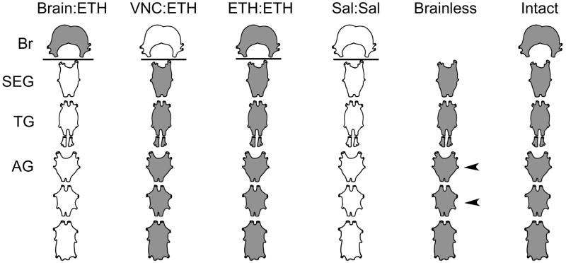
Ecdysis, or the shedding of the old cuticle, depends on coordinated stereotyped behaviors, regulated by a number of neuropeptides. In the hornworm, Manduca sexta, two neuropeptides interact, namely ecdysis-triggering hormone (ETH) and eclosion hormone. We looked at the effects of ETH in vivo and in vitro, on the brain and the ventral nerve cord to determine the roles played by these hormones. We monitored ecdysis onset and the presence of cGMP and eclosion hormone immunoreactivity. In vivo, only a fraction of larvae lacking the cell bodies containing eclosion hormone, and injected with ETH, were able to undergo ecdysis, with a delayed response. These animals showed strongest cGMP immunoreactivity in the subesophageal and thoracic ganglia, with concomitant reductions in eclosion hormone immunoreactivity in descending axons in comparison with animals not undergoing ecdysis. Animals lacking the brain showed reduced to no cGMP levels in all ganglia. In vitro, isolated CNS preparations lacking the brain initiated ecdysis motor programs after incubation in ETH, with faster onset times than controls, and with reduced cGMP immunoreactivity. If ETH was applied only to the brain of the isolated CNS, cGMP immunoreactivity was noted primarily in the subesophageal and thoracic ganglia, with a decrease in eclosion hormone immunoreactivity in descending axons. ETH addition to the rest of the nerve cord showed reduced eclosion hormone immunoreactivity but little to no cGMP immunoreactivity in any ganglion. Controls showed strong cGMP immunoreactivity in all ganglia, and even greater reductions in eclosion hormone staining after ETH application. These results support previous suggestions that eclosion hormone is required for a positive feedback loop with ETH as well as onset of an inhibitory component, but also suggest that ETH stimulates eclosion hormone release at multiple spike initiation zones. The resultant up regulation of cGMP does not appear to be required for onset of ecdysis. A new model for ecdysis regulation is considered.


Figures

Fig. 1
Fig. 1
Model for the regulation of ecdysis in the larval CNS of M. sexta. A description of the model is provided in the introduction. Abbreviations: (Br) brain; (SEG/TG) subesophageal and thoracic ganglia; (AG) abdominal ganglia; (PETH) Pre-Ecdysis Triggering Hormone; (ETH) Ecdysis Triggering Hormone; (EH) Eclosion Hormone; (?) unknown cell phenotype; (CCAP) Crustacean Cardioactive Peptide; (MIP) MyoInhibitory Peptide; (DH) Diuretic Hormone; (cGMPir) cGMP immunoreactivity; (CPG) central pattern generator; (Pre I) pre-ecdysis I; (Pre II) pre-ecdysis II; (E) ecdysis; (post) post-ecdysis. Dashed and solid arrows show excitatory effects. Capped line shows an inhibitory effect. Dashed line depicts separation of ganglia. dark cells have been shown to have increases in cGMPir. Clear cells have not. Numbers are discussed in the introduction.
Fig. 2
Fig. 2
Schematic representation of the isolated CNS preparations used for in vitro Vaseline dam experiments (Brain:ETH, VNC:ETH, ETH:ETH; Sal:Sal) and electrophysiology (Brainless and Intact). The CNS is truncated to show the brain (Br), subesophageal ganglion (SEG), one thoracic ganglion (TG) and 3 abdominal ganglia (AG) with connectives intact. Grey shaded tissues represent sites of ETH application and white tissues represent sites of saline application. The lines show the sites of the Vaseline dam. The arrowheads identify the sites of suction electrode placement for electrophysiological recordings. (VNC): Ventral nerve cord; (Sal): Saline.
Fig. 3
Fig. 3
EH immunoreactivity in axons of the terminal abdominal ganglia from ecdysing and non-ecdysing animals. Examples of confocal stacked images of EH-immunoreactive axons are depicted from (A) sham-operated saline-injected “sham (saline)” and (B) sham-operated ETH-injected “sham (ETH)” animals. Sham (saline) animals did not go through ecdysis, while sham (ETH) animals did. Scale bar = 0.01 mm. (C) Relative pixel intensities of EH-immunoreactive axons from control and experimental groups. Sham: sham brain surgery; no MBr (E): removal of midbrain, and animals still ecdysed; no MBr (no E): removal of midbrain, and animals did not ecdyse after 90 min. Asterisks denote significant differences (P<0.05), using Tukey’s HSD on transformed data after One-way Analysis of Variance.
Fig. 4
Fig. 4
Percentage of tissues stained for cGMP immunoreactivity in the (A) IN704 interneurons or (B) NS27 neurons of the subesophageal ganglion (SEG), thoracic ganglia (TG) or abdominal ganglia (AG) when incubated with ETH for 40 minutes. Intact or brainless preparations were incubated with 1 μM ETH. Letters are significantly different from controls (p<0.05). Sample sizes ranged from 26–28 tissues for each group.
Fig. 5
Fig. 5
Representative photomicrographs of cGMP immunoreactivity in isolated CNS preparations with a Vaseline dam between the brain and VNC, after incubated in 1 μM ETH for 40 min. (A) Entire CNS incubated in ETH (ETH:ETH); (B) Brain incubated in ETH and VNC incubated in saline (Brain:ETH); (C) Brain incubated in saline and VNC incubated in ETH (VNC:ETH). Subesophageal ganglion (SEG); Thoracic ganglion (TG); Abdominal ganglia (AG); Terminal abdominal ganglion (TAG). Asterisks indicate cells that were not scored for cGMP immunoreactivity. Scale bar = 150 μm.
Fig. 6
Fig. 6
Percentage of tissues stained for cGMP immunoreactivity in the (A) IN704 interneurons or (B) NS27 neurons of the subesophageal ganglion (SEG), thoracic ganglia (TG) or abdominal ganglia (AG) when incubated with 1 μM ETH for 40 minutes. Tissues received ETH or saline on either side of a Vaseline dam between the brain and VNC. Controls received ETH on both sides of the dam (ETH:ETH). Other groups received ETH on the brain alone (Brain:ETH) or the VNC alone (VNC:ETH). Letters represent similarities between or within groups (p>0.05). Sample sizes ranged from 26–28 tissues for each group.
Fig. 7
Fig. 7
Average relative pixel intensity of all cells showing cGMP immunoreactivity in the (A) IN704 interneurons or (B) NS27 neurons of the subesophageal ganglion (SEG), thoracic ganglia (TG) or abdominal ganglia (AG) when incubated with 1 μM ETH for 40 minutes. Tissues received ETH or saline on either side of a Vaseline dam between the brain and VNC. Controls received ETH on both sides of the dam (ETH:ETH). Other groups received ETH on the brain alone (Brain:ETH) or the VNC alone (VNC:ETH). Letters represent similarities between or within groups (p>0.05). Sample sizes ranged from 26–28 tissues for each group.
Fig. 8
Fig. 8
EH immunoreactivity in axons of the terminal abdominal ganglia of isolated CNS preparations with various saline or ETH treatments. (A, B) Representative confocal stacked images of EH-immunoreactive axons after incubation in saline or ETH, respectively. Arrows show stained axons. Scale bar = 0.01 mm. (C) Relative pixel intensities of axons of the terminal abdominal ganglia of isolated CNS preparations. Treatments are noted below each bar. [Sal:Sal] saline applied to the entire CNS (brain and VNC); [ETH:ETH] ETH applied to the entire CNS (brain and VNC); [BR:ETH] ETH applied to the brain alone, with saline on the VNC; [VNC:ETH] ETH applied to the entire VNC, but not the brain, with saline applied to the brain. Letters denote significant differences (P<0.05), using Tukey’s HSD on transformed data after One-way Analysis of Variance. Sample sizes are noted in white.
Fig. 9
Fig. 9
Updated model for the regulation of ecdysis in the larval CNS. See Fig. 1 caption for details on abbreviations. The behavioral repertoire has been divided into two parts: beginning sequence of behaviors (A), and end sequence of behaviors (B).

Similar articles

Cited by

References

    1. Adams ME, Zitnan D. Identification of ecdysis-triggering hormone in the silkworm Bombyx mori. Biochem Biophys Res Commun. 1997;230:188–191. - PubMed
    1. Calabrese RL. Control of multiple impulse-initiation sites in a leech intemeuron. J Neurophysiol. 1980;44:878–896. - PubMed
    1. Clark A, del Campo M, Ewer J. Neuroendocrine Control of Larval Ecdysis Behavior in Drosophila: Complex Regulation by Partially Redundant Neuropeptides. J Neurosci. 2004;24(17):4283–4292. - PMC - PubMed
    1. Copenhaver PF, Truman JW. The role of eclosion hormone in the larval ecdyses of Manduca Sexta. J Insect Physiol. 1982;28:695–701.
    1. Davis NT, Homberg U, Dircksen H, Levine RB, Hildebrand JG. Crustacean cardioactive peptide-immunoreactive neurons in the hawkmoth Manduca sexta and changes in their immunoreactivity during postembryonic development. J Comp Neurol. 1993;338:612–627. - PubMed

Publication types