Review
. 2009 Aug;20(4):398-404.
doi: 10.1016/j.copbio.2009.07.007. Epub 2009 Aug 21.

Generation of new protein functions by nonhomologous combinations and rearrangements of domains and modules

Affiliations
Review

Generation of new protein functions by nonhomologous combinations and rearrangements of domains and modules

Shohei Koide. Curr Opin Biotechnol. 2009 Aug.

Abstract

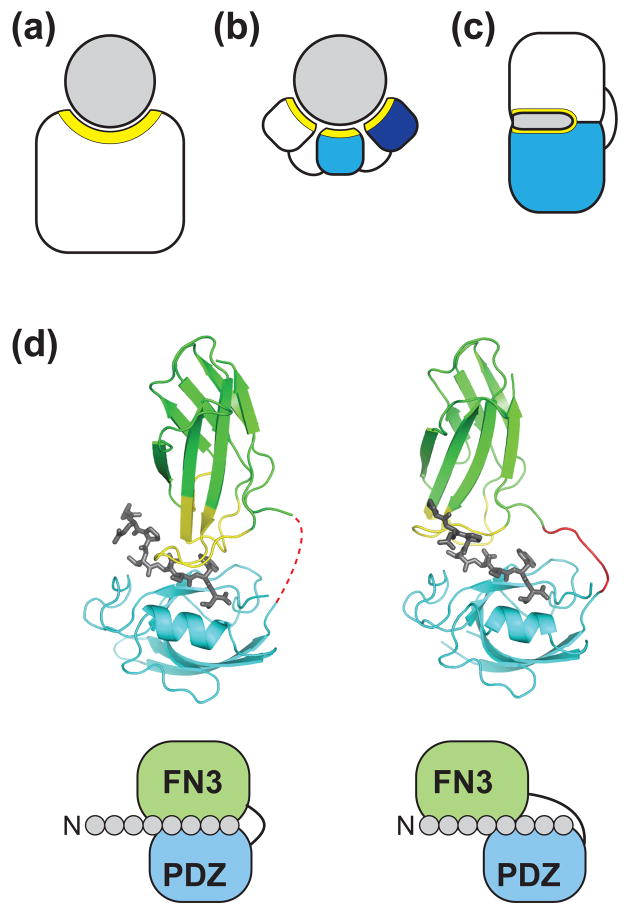
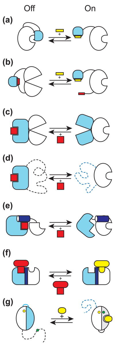
Generation of novel protein functions is a major goal in biotechnology and also a rigorous test for our understanding of the relationship between protein structure and function. Early examples of protein engineering focused on design and directed evolution within the constraints of the original protein architecture, exemplified by the highly successful fields of antibody and enzyme engineering. Recent studies show that protein engineering strategies which step away from these natural architectures, that is by manipulating the organization of domains and modules thus mimicking nonhomologous recombination, are highly effective in producing complex and sophisticated functions in terms of both molecular recognition and regulation.


Figures

Figure 1
Figure 1
Generation of new molecular recognition functions by combining domains. (a–c) Schematic drawing of different types of binding protein architectures. (a) "Single-patch" recognition by a conventional binding protein. (b) Multivalent (multi-epitope) recognition by multiple modules linked with flexible linkers. (c) Synergistic recognition of a contiguous target surface by a single binding site constructed at the interface of two domains. The target (or antigen) is colored in gray. Recognition surfaces of the binding proteins are marked in yellow. (d) Crystal structures of two affinity clamps (PDB ID: 2QBW and 3CH8) showing the ability of directed domain-interface evolution to produce diverse interface configurations. The schemes below the structural models show simplified views of the interactions between the 8-residue target peptide and the affinity clamps. The molecular graphics was generated using PyMOL (www.pymol.org).
Figure 2
Figure 2
Molecular mechanisms used to create synthetic regulatory circuits and sensors by module/domain recombination and rearrangement. The input module is shown in light blue and the output module is in white. The output module is in the "on" state on the right side and "off" on the left. An activator is shown in yellow and an inhibitor in red. (a) Steric allostery. The input module physically occludes the active site of the output module, which can be relieved by a ligand (yellow) for the input module. (b) Conformational allostery. Interactions involving the input module and its intramolecular ligand distort the conformation of the output module. A ligand in trans removes the inhibition. (c) Conformational coupling. Two modules are connected in such a way that only one of them is in the active conformation at a given time. A ligand binding to the active state of the input module inhibits the output module. (d) Mutually exclusive folding. The dashed lines indicate unfolded segments. This is an extreme case of conformational coupling depicted in panel c. (e) Segmental overlap creating mutually exclusive folding of a shared helix (shown in dark blue) and thus conformational allostery. A ligand for the input module stabilizes docking of the shared helix to the rest of the input module and thus inactivates the output module. (f) Segmental overlap creating steric allostery. Two modules are fused in such a way that a ligand to the input module (red) physically occludes binding of a ligand to the output module (yellow). (g) Alternative folding frames for creating a sensor. The blue and gray portions denote equivalent segment of the starting protein that has been duplicated. The blue segment is mutated so that it no longer binds to a ligand but it interacts with the rest of the protein more strongly than the gray segment thereby favoring the inactive state (the left side of the arrows). A ligand shifts the equilibrium to the right, which changes the distance between fluorophores (the yellow and green circles). The schemes (a) and (b) are made after Dueber et al. [31], (e) after Strickland et al. [44] and (g) after Stratton et al. [48].

Similar articles

Cited by

References

    1. Pluckthun A, Mayo SL. The design of evolution and the evolution of design. Curr Opin Struct Biol. 2007;17:451–453.
    1. Kortemme T, Baker D. Computational design of protein-protein interactions. Curr Opin Chem Biol. 2004;8:91–97. - PubMed
    1. Winter G, Griffiths AD, Hawkins RE, Hoogenboom HR. Making antibodies by phage display technology. Annu Rev Immunol. 1994;12:433–455. - PubMed
    1. Binz HK, Amstutz P, Pluckthun A. Engineering novel binding proteins from nonimmunoglobulin domains. Nat Biotechnol. 2005;23:1257–1268. - PubMed
    1. Looger LL, Dwyer MA, Smith JJ, Hellinga HW. Computational design of receptor and sensor proteins with novel functions. Nature. 2003;423:185–190. - PubMed

Publication types