Procoagulant alveolar microparticles in the lungs of patients with acute respiratory distress syndrome
- PMID: 19700643
- PMCID: PMC2793184
- DOI: 10.1152/ajplung.00214.2009
Procoagulant alveolar microparticles in the lungs of patients with acute respiratory distress syndrome
Abstract
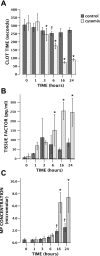
Coagulation and fibrinolysis abnormalities are observed in acute lung injury (ALI) in both human disease and animal models and may contribute to ongoing inflammation in the lung. Tissue factor (TF), the main initiator of the coagulation cascade, is upregulated in the lungs of patients with ALI/acute respiratory distress syndrome (ARDS) and likely contributes to fibrin deposition in the air space. The mechanisms that govern TF upregulation and activation in the lung are not well understood. In the vascular space, TF-bearing microparticles (MPs) are central to clot formation and propagation. We hypothesized that TF-bearing MPs in the lungs of patients with ARDS contribute to the procoagulant phenotype in the air space during acute injury and that the alveolar epithelium is one potential source of TF MPs. We studied pulmonary edema fluid collected from patients with ARDS compared with a control group of patients with hydrostatic pulmonary edema. Patients with ARDS have higher concentrations of MPs in the lung compared with patients with hydrostatic edema (25.5 IQR 21.3-46.9 vs. 7.8 IQR 2.3-27.5 micromol/l, P = 0.009 by Mann-Whitney U-test). These MPs are enriched for TF, have procoagulant activity, and likely originate from the alveolar epithelium [as measured by elevated levels of RAGE (receptor for advanced glycation end products) in ARDS MPs compared with hydrostatic MPs]. Furthermore, alveolar epithelial cells in culture release procoagulant TF MPs in response to a proinflammatory stimulus. These findings suggest that alveolar epithelial-derived MPs are one potential source of TF procoagulant activity in the air space in ARDS and that epithelial MP formation and release may represent a unique therapeutic target in ARDS.
Figures


Comment in
-
Thinking small, but with big league consequences: procoagulant microparticles in the alveolar space.Am J Physiol Lung Cell Mol Physiol. 2009 Dec;297(6):L1033-4. doi: 10.1152/ajplung.00335.2009. Epub 2009 Oct 2. Am J Physiol Lung Cell Mol Physiol. 2009. PMID: 19801449 No abstract available.
References
-
- American College of Chest Physicians/Society of Critical Care Medicine Consensus Conference Definitions for sepsis and organ failure and guidelines for the use of innovative therapies in sepsis. Crit Care Med 20: 864– 874, 1992 - PubMed
-
- Abid Hussein MN, Meesters EW, Osmanovic N, Romijn FP, Nieuwland R, Sturk A. Antigenic characterization of endothelial cell-derived microparticles and their detection ex vivo. J Thromb Haemost 1: 2434– 2443, 2003 - PubMed
-
- Aras O, Shet A, Bach RR, Hysjulien JL, Slungaard A, Hebbel RP, Escolar G, Jilma B, Key NS. Induction of microparticle- and cell-associated intravascular tissue factor in human endotoxemia. Blood 103: 4545– 4553, 2004 - PubMed
Publication types
MeSH terms
Substances
Grants and funding
LinkOut - more resources
Full Text Sources
Other Literature Sources
Miscellaneous

