. 2009 Sep;175(3):1338-47.
doi: 10.2353/ajpath.2009.090234. Epub 2009 Aug 21.

A key role for the integrin alpha2beta1 in experimental and developmental angiogenesis

Affiliations

A key role for the integrin alpha2beta1 in experimental and developmental angiogenesis

James D San Antonio et al. Am J Pathol. 2009 Sep.

Abstract

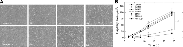
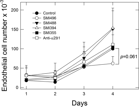
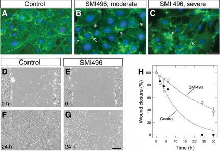
The alpha2beta1 integrin receptor plays a key role in angiogenesis. Here we investigated the effects of small molecule inhibitors (SMIs) designed to disrupt integrin alpha2 I or beta1 I-like domain function on angiogenesis. In unchallenged endothelial cells, fibrillar collagen induced robust capillary morphogenesis. In contrast, tube formation was significantly reduced by SMI496, a beta1 I-like domain inhibitor and by function-blocking anti-alpha2beta1 but not -alpha1beta1 antibodies. Endothelial cells bound fluorescein-labeled collagen I fibrils, an interaction specifically inhibited by SMI496. Moreover, SMI496 caused cell retraction and cytoskeletal collapse of endothelial cells as well as delayed endothelial cell wound healing. SMI activities were examined in vivo by supplementing the growth medium of zebrafish embryos expressing green fluorescent protein under the control of the vascular endothelial growth factor receptor-2 promoter. SMI496, but not a control compound, interfered with angiogenesis in vivo by reversibly inhibiting sprouting from the axial vessels. We further characterized zebrafish alpha2 integrin and discovered that this integrin is highly conserved, especially the I domain. Notably, a similar vascular phenotype was induced by morpholino-mediated knockdown of the integrin alpha2 subunit. By live videomicroscopy, we confirmed that the vessels were largely nonfunctional in the absence of alpha2beta1 integrin. Collectively, our results provide strong biochemical and genetic evidence of a central role for alpha2beta1 integrin in experimental and developmental angiogenesis.


Figures

Figure 1
Figure 1
Structure of small molecule inhibitors of the α2β1 integrin receptor. Low Mr (<600 Da) benzene scaffold derivatives designed to bind and functionally disrupt the α2β1 integrin receptor. Inhibitors and their receptor target sites include the following: SMI355, 394, and 418, α2I domain; SMI488 and 723 (not shown) negative controls; and SMI496, β1I-like domain.
Figure 2
Figure 2
Disruption of collagen-induced in vitro capillary morphogenesis by SMI496, an inhibitor of α2β1-integrin function. A: Photomicrographs of live endothelial cells undergoing in vitro capillary morphogenesis in serum-free defined media supplemented with VEGF-fibroblast growth factor-2 (5 ng/ml each) ± SMI496 (100 nmol/L). Note the complete block of capillary-like formation by SMI496, clearly visible at 24 hours. Similar results were obtained with an anti-α2β1 integrin function-blocking antibody (5 μg/ml). No effect was observed with any of the other SMIs (not shown). Scale bar = ∼100 μm. B: Quantification of three independent experiments similar to that shown in A. Each experiment was run in triplicate. Values represent the mean ± SE. Notice that only SMI496 is capable of blocking capillary formation (***P < 0.001). A similar inhibition was also evoked by an anti-α2β1 integrin function-blocking antibody. Angiogenic areas were measured using the ImageJ program.
Figure 3
Figure 3
Effects of SMIs on growth. Notice the lack of growth inhibition by all of the SMIs tested. By 4 days a borderline (P = 0.061) inhibition of cell growth by SMI496 was observed. Values represent the mean ± SE of three independent experiments run in triplicate. Endothelial cells were supplemented daily with fresh media containing the various SMIs (all at ∼100 nmol/L) or 10 μg/ml of an anti-α2β1 integrin function-blocking antibody. On each day, plates were fixed with glutaraldehyde and on day 4, all cultures were stained with crystal violet, the dye was detergent extracted, and cell number was determined spectrophotometrically.
Figure 4
Figure 4
Effects of SMIs on endothelial cell-collagen fibril ligation in suspension. Note that SMI496 but not the negative compound SMI723 disrupts FITC-labeled collagen binding. The anti-α2β1 integrin function-blocking antibody but not an anti-CD2 blocking antibody has a similar effect. For flow cytometry studies, endothelial cells were trypsinized, allowed to recover for 2 hours, and then exposed to SMIs (50 nmol/L) or antibodies (100 μg/ml) in PBS ± 1% bovine serum albumin. FITC-collagen (50 μg/ml) was then added. Cell-FITC collagen interactions were analyzed on a Beckman Coulter EPICS flow cytometer. Anti-CD2 antibody and the negative control SMI723 were used to obtain baseline measures of FITC-collagen staining. Values represent the mean ± SE from three independent experiments run in triplicate (***P < 0.001, by Student’s t-test). Scatterplots are found in Supplemental Figure S2, see http://ajp.amipathol.org.
Figure 5
Figure 5
Effects of SMIs on endothelial cell attachment to collagen substrata and migration. Fluorescent micrographs of endothelial cells cultured on type I collagen treated for 30 minutes with either vehicle (A) or SMI496 (100 nmol/L) (B and C). The arrows point to the collapse and coalescence of actin bundles. The asterisks point to areas of endothelial cell retraction. The cells were fixed, detergent-permeabilized, and stained with phalloidin-FITC (green) to visualize actin stress fibers and counterstained with DAPI (blue) to visualize nuclei. Note the partial collapse of the actin cytoskeleton (arrows, B and C). The experiments were repeated three times with similar results. Scale bar = 20 μm. D–G: Light micrographs of “wounded” endothelial monolayers and the effect of SMI496. Confluent endothelial cell layers on collagen films were scraped with a 200-μl pipette tip. The cells were rinsed with PBS and resupplemented with either media alone or media supplemented with SMI496 (100 nmol/L). Micrographs were taken at various time points. Scale bar = 100 μm. H: Quantification of endothelial cell wound closure over time. The areas within the scratches were quantified using ImageJ software and expressed as percentage of wound closure at time 0. Values represent the mean ± SE of two independent experiments run in triplicate.
Figure 6
Figure 6
SMI496 inhibits angiogenesis in the zebrafish trunk. A–C: Bright-field and fluorescent micrographs of live vegfr2-gfp zebrafish controls at 2 dpf. Note the proper formation of the major axial vessels, dorsal aorta (DA), and posterior cardinal vein (PCV), as well as the inter-segmental vessels (ISVs) and the dorsal longitudinal anastomotic vessel (DLAV). D–I: Representative embryos treated from 7 hpf to 2 dpf with SMI496 at the indicated concentrations. Note the abnormal and stunted formation of the intersegmental vessels (arrows in F and I) and the absence of the dorsal longitudinal anastomotic vessel. J–L: Representative images of partial recovery after removal of SMI496 from the embryo medium and subsequent growth for an additional 3 days in compound-free water. M–O: Representative fluorescent images of live vegfr2-gfp transgenic zebrafish at 2 dpf treated with SMI355 at the designated concentrations for 4 hours starting at 7 hpf. Note normal vasculogenesis and angiogenesis. All panels are oriented with rostral to the left and all lateral views are from the left side. Scale bars = ∼300 μm.
Figure 7
Figure 7
Characterization of the α2 morphant phenotype. A: Representative bright-field image of four 1.5 dpf control embryos. B: Representative bright-field image of six matched α2-MO injected embryos. C: Control vasculature as visualized by GFP and corresponding to the embryos presented in A. D: α2 morphant vasculature corresponding to the embryos presented in B. Note the defect in trunk and tail angiogenesis. E: Summary of the dose-response relationship between morpholino injection amount and overall phenotypic penetrance. Scale bar = 1 mm. α2 knockdown inhibits multiple aspects of developmental angiogenesis. F–I: Fluorescent micrographs of live vegfr2-gfp zebrafish embryos focusing on the vasculature at 1.5 dpf of control and integrin α2 morphant embryos. F: Representative control trunk to tail region exhibiting complete formation of the axial vessels, ISVs and caudal venous plexus (CV). G and H: morphant trunk regions exhibiting abnormal ISV development. I: Representative morphant tail region exhibiting abnormal CV development. Scale bars = ∼200 μm.
Figure 8
Figure 8
α2 knockdown disrupts angiogenic subintestinal vessel (SIV) development. A: Representative control embryo displaying clear formation of the subintestinal vessels by 3 dpf. B–D: Matched α2 morphant embryos exhibiting inhibition of subintestinal vessel formation in a dose-dependent manner. All vessels were visualized by endogenous alkaline phosphatase staining. Scale bar = 250 μm. E: Graphic summary of the number of subintestinal vessel plexus intersection points. Data are the mean ± SD of 10 embryos/dose from two independent experiments.

Similar articles

Cited by

References

    1. Folkman J. Role of angiogenesis in tumor growth and metastasis. Semin Oncol. 2002;29:15–18. - PubMed
    1. Davis GE, Senger DR. Endothelial extracellular matrix: biosynthesis, remodeling, and functions during vascular morphogenesis and neovessel stabilization. Circ Res. 2005;97:1093–1107. - PubMed
    1. Davis GE, Senger DR. Extracellular matrix mediates a molecular balance between vascular morphogenesis and regression. Curr Opin Hematol. 2008;15:197–203. - PubMed
    1. Abair TD, Sundaramoorthy M, Chen D, Heino J, Ivaska J, Hudson BG, Sanders CR, Pozzi A, Zent R. Cross-talk between integrins α1β1 and α2β1 in renal epithelial cells. Exp Cell Res. 2008;314:3593–3604. - PMC - PubMed
    1. Senger DR, Claffey KP, Benes JE, Perruzzi CA, Sergiou AP, Detmar M. Angiogenesis promoted by vascular endothelial growth factor: regulation through α1β1 and α2β1 integrins. Proc Natl Acad Sci USA. 1997;94:13612–13617. - PMC - PubMed

Publication types

MeSH terms

LinkOut - more resources