. 2009 Oct;37(18):6092-104.
doi: 10.1093/nar/gkp674. Epub 2009 Aug 21.

Identification and functional characterization of two new transcriptional variants of the human p63 gene

Affiliations

Identification and functional characterization of two new transcriptional variants of the human p63 gene

Marina Mangiulli et al. Nucleic Acids Res. 2009 Oct.

Abstract

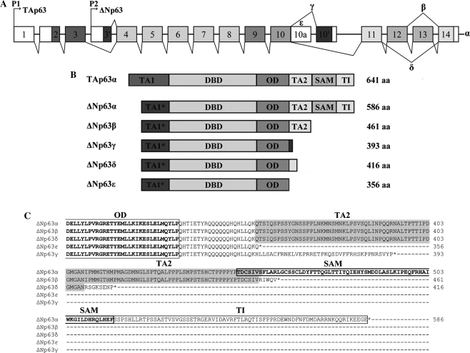
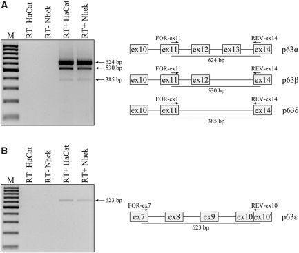
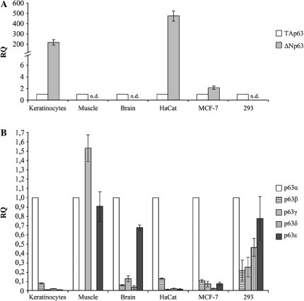
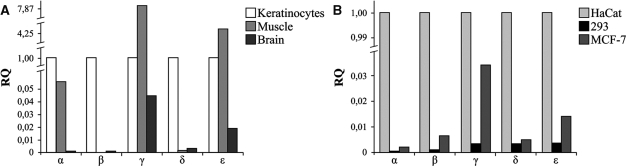
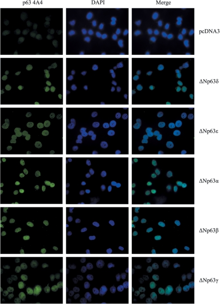
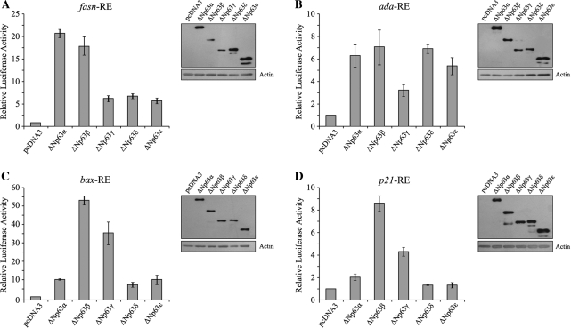
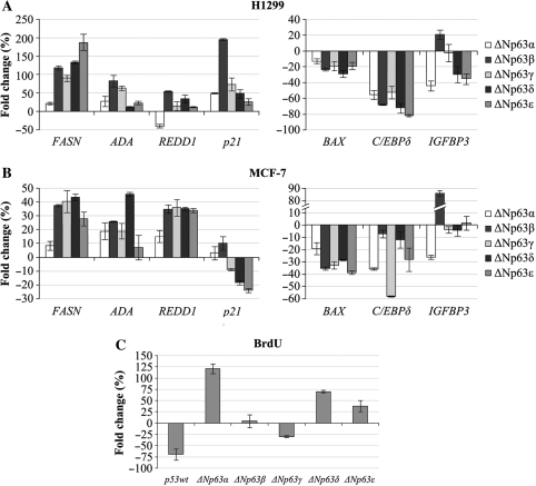
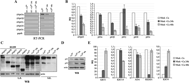
p63 belongs to a family of transcription factors, which, while demonstrating striking conservation of functional domains, regulate distinct biological functions. Its principal role is in the regulation of epithelial commitment, differentiation and maintenance programs, during embryogenesis and in adult tissues. The p63 gene has a complex transcriptional pattern, producing two subclasses of N-terminal isoforms (TA and DeltaN) which are alternatively spliced at the C-terminus. Here, we report the identification of two new C-terminus p63 variants, we named p63 delta and epsilon, that increase from 6 to 10 the number of the p63 isoforms. Expression analysis of all p63 variants demonstrates a tissue/cell-type-specific nature of p63 alternative transcript expression, probably related to their different cellular functions. We demonstrate that the new p63 variants as DeltaN isoforms are active as transcription factors as they have nuclear localization and can modulate the expression of p63 target genes. Moreover, we report that, like DeltaNp63alpha, DeltaNp63delta and epsilon sustain cellular proliferation and that their expression decreases during keratinocyte differentiation, suggesting their involvement in this process. Taken together, our results demonstrate the existence of novel p63 proteins whose expression should be considered in future studies on the roles of p63 in the regulation of cellular functions.


Figures

Figure 1.
Figure 1.
Human p63 gene. (A) Schematic representation of human p63 gene structure: alternative promoters (P1 and P2), previously identified alternative splicing events (α, β and γ) and novel events (δ and ϵ) are indicated. (B) ΔNp63 protein isoforms are reported with their molecular size. TA1*: TA of ΔN isoforms; DBD: DNA-binding domain; OD: oligomerization domain; TA2: C-terminal TA; SAM: sterile alpha motif; TI: inhibitory domain. (C) Amino acid alignment among the C-terminal regions of δ and ε p63 proteins and the known α, β and γ.
Figure 2.
Figure 2.
In vivo identification of novel p63 δ and ϵ transcripts. (A) RT–PCR analysis of HaCat and Nhek RNAs which identifies the α, β and δ transcripts; on the right, a schematic representation of the portion of p63 transcripts amplified with the expected size. (B) RT–PCR analysis from HaCat and Nhek RNAs which identifies the ε transcripts; on the right, a schematic representation of the portion of p63 transcripts amplified with the expected size. Primers used are indicated by arrows.
Figure 3.
Figure 3.
Identification of the novel ΔNp63δ and ϵ proteins. Endogenous p63 protein expression was evaluated by western analysis in HaCat and MCF-7 cells. Five micrograms of lysate of H1299 over-expressing ΔNp63α, β, γ, δ and ϵ proteins were used as molecular marker to assess an identity to each ΔNp63 isoform in 60 μg of Hacat and MCF-7 lysates. The pan-p63 4A4 antibody was used to detect ΔNp63 isoforms. Ectopically expressed ΔNp63α, β and γ show a higher molecular mass than expected as they express the myc-tag in their N-terminus. Arrows on the right indicate the position of ΔNp63δ and ϵ isoforms in HaCat and MCF-7 lysate. The molecular weights of protein markers are shown on the left.
Figure 4.
Figure 4.
Expression profile of human p63 transcripts in different normal tissues and cell lines. (A) TA and ΔN p63 transcripts levels were quantified by qRT–PCR. Values are expressed as fold change respect to TA transcripts, used as calibrator, after internal normalization for gapdh expression (see ‘Material and Methods’ section). (B) p63 α, β, γ, δ and ϵ mRNA levels were quantified by qRT–PCR. Values are expressed as fold change respect to α variant, used as calibrator, after internal normalization to gapdh expression.
Figure 5.
Figure 5.
Differential expression of human p63 transcripts among different normal tissues (A) and cell lines (B). mRNA levels of p63 variants (α, β, γ, δ and ϵ) were quantified by qRT–PCR. Values are expressed as fold change respect to keratinocytes and HaCat cells, used as calibrators, respectively for tissues and cell lines, after internal normalization to gapdh expression.
Figure 6.
Figure 6.
Nuclear localization of ΔNp63δ and ΔNp63ε isoforms. H1299 cells were transiently transfected for 24 h with individual vectors expressing the novel ΔNp63δ and ϵ isoforms and ΔNp63α, β and γ isoforms as controls, and immunostained using the p63 4A4 antibody and a FITC-conjugated secondary antibody. Nuclei were counterstained with DAPI.
Figure 7.
Figure 7.
ΔNp63δ and ΔNp63ε isoforms have transactivation activity. H1299 cells were transiently co-transfected with the pcDNA3 empty vector or expressing ΔNp63 α, β, γ, δ and ϵ and the reporter constructs pGL3-fasnRE (A), pGL3-adaRE (B), pGL3-baxRE (C) and pGL3-p21RE (D). The fold increase in relative luciferase activity was calculated using the empty pcDNA3 vector as control. The results represent the average of at least three independent experiments and are shown with the standard deviations. For each reporter construct, the expression level of the ΔNp63 isoforms was checked by western blotting.
Figure 8.
Figure 8.
Ectopic expression of ΔNp63δ and ΔNp63ε isoforms affects the in vivo expression of p63 target genes and promotes cellular proliferation. H1299 (A) and MCF-7 (B) cell lines were transfected with the pcDNA3 vector or vector expressing ΔNp63 α, β, γ, δ and ϵ and harvested after 48 h. mRNA levels of fasn, ada, redd1, p21, bax, C/EBPdelta and igfbp-3 genes were quantified by qRT–PCR. Values are expressed as percentage of fold change respect to the control (pcDNA3), after internal normalization to gapdh expression. (C) Bromodeoxyuridine (BrdU) incorporation after 3 h pulses by MCF-7 cells transfected with pcDNA3 control vector, or with the indicated expression vectors.
Figure 9.
Figure 9.
Expression of ΔNp63δ and ΔNp63ε isoforms is modulated during keratinocyte differentiation. Differentiation of proliferating keratinocytes was induced by varying the extracellular Ca2+ concentration from 0 to 2 mM and the medium serum concentration from 1× to 0.1× for 24 and 48 h. (A) RT–PCR analysis of full-length ΔNp63 mRNA levels in proliferating keratinocytes (–Ca2+) and at 24 and 48 h after Ca2+ addition. (B) qRT–PCR analyses of ΔNp63 variants in proliferating keratinocytes (–Ca2+) and at 24 h and 48 h after Ca2+ addition. Values are expressed as fold change respect to the control (–Ca2+) after internal normalization to gapdh expression. (C) Western blotting analysis of all ΔNp63 proteins in proliferating keratinocytes (–Ca2+) and at 24 and 48 h after Ca2+ addition. Ectopically expressed ΔNp63 proteins were used as molecular marker to assess an identity to each ΔNp63 isoform. The pan-p63 4A4 antibody was used to detect p63 isoforms. L.E. = long exposure, S.E. = short exposure. (D) Western blotting analysis of p21 and p53, used as controls of the differentiation induction. (E) qRT–PCR analysis of ada, redd1, keratin 4, keratin 14 and keratin 1 mRNA levels during keratinocyte differentiation. Values are expressed as fold induction respect to the control (–Ca2+), after internal normalization to gapdh expression.

References

    1. Yang A, Kaghad M, Wang Y, Gillett E, Fleming MD, Dotsch V, Andrews NC, Caput D, McKeon F. p63, a p53 homolog at 3q27-29, encodes multiple products with transactivating, death-inducing, and dominant-negative activities. Mol. Cell. 1998;2:305–316. - PubMed
    1. Helton ES, Zhu J, Chen X. The unique NH2-terminally deleted (DeltaN) residues, the PXXP motif, and the PPXY motif are required for the transcriptional activity of the DeltaN variant of p63. J. Biol. Chem. 2006;281:2533–2542. - PubMed
    1. Ghioni P, Bolognese F, Duijf PH, Van Bokhoven H, Mantovani R, Guerrini L. Complex transcriptional effects of p63 isoforms: identification of novel activation and repression domains. Mol. Cell Biol. 2002;22:8659–8668. - PMC - PubMed
    1. Serber Z, Lai HC, Yang A, Ou HD, Sigal MS, Kelly AE, Darimont BD, Duijf PH, Van Bokhoven H, McKeon F, et al. A C-terminal inhibitory domain controls the activity of p63 by an intramolecular mechanism. Mol. Cell Biol. 2002;22:8601–8611. - PMC - PubMed
    1. Donehower LA, Harvey M, Slagle BL, McArthur MJ, Montgomery CA, Jr, Butel JS, Bradley A. Mice deficient for p53 are developmentally normal but susceptible to spontaneous tumours. Nature. 1992;356:215–221. - PubMed

Publication types

MeSH terms

Associated data