

Prevalence of oral lesions among Saudi dental patients
- PMID: 19700894
- PMCID: PMC3290046
- DOI: 10.4103/0256-4947.55166
Prevalence of oral lesions among Saudi dental patients
Abstract
Background and objectives: Few studies have been conducted in the Saudi population on oral mucosal lesions. The purpose of this study was to evaluate the type and extent of oral lesions in a study among dental patients at a college of dentistry in Saudi Arabia.
Patients and methods: Over a 3-year period, 2552 dental outpatients were interviewed and investigated clinically for the presence of oral mucosal conditions. A thorough oral clinical examination was performed, including a radiographic examination. The diagnosis was confirmed histopathologically when necessary.
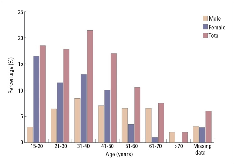
Results: Of 383 (15.0%) patients found to have oral mucosal lesions, females constituted 57.7% (n=221) and males 42.3% (n=162). The age range of the patients was between 15 to 73 years with a mean age of 38.2 years. The most commonly affected age group was 31 to 40 years, which comprised 21.4% (n=82) of all affected individuals. The least affected age group were individuals older than 61 years. The most common lesion was Fordyce granules (3.8%; n=98), followed by leukoedema (3.4%; n=86) and traumatic lesions (ulcer, erosion) in 1.9% (n=48). Tongue abnormalities were present in 4.0% (n=101) of all oral conditions observed, ranging from 1.4% (n=36) for fissured tongue to 0.1% (n=2) for bifid tongue. Other findings detected were torous platinus (1.3%; n=34), mandibular tori (0.1%; n=2) aphthous ulcer (0.4%; n=10), herpes simplex (0.3%; n=7), frictional hyperkeratosis (0.9%; n=23), melanosis (0.6%; n=14), lichen planus (0.3%; n=9) and nicotinic stomatitis (0.5%; n=13).
Conclusion: The findings of this study provide information on the types and prevalence of oral lesions among Saudi dental patients. This provides baseline data for future studies about the prevalence of oral lesions in the general population.
Figures
Similar articles
-
Prevalence and distribution of oral mucosal non-malignant lesions in the western Sicilian population.Minerva Stomatol. 2016 Aug;65(4):191-206. Minerva Stomatol. 2016. PMID: 27374359
-
Prevalence of potentially malignant oral mucosal lesions among tobacco users in Jeddah, Saudi Arabia.Asian Pac J Cancer Prev. 2014;15(2):757-62. doi: 10.7314/apjcp.2014.15.2.757. Asian Pac J Cancer Prev. 2014. PMID: 24568491
-
Oral mucosal lesions in a representative cross-sectional study of aging Germans.Community Dent Oral Epidemiol. 2000 Oct;28(5):390-8. doi: 10.1034/j.1600-0528.2000.028005390.x. Community Dent Oral Epidemiol. 2000. PMID: 11014516
-
Epidemiology of the most common oral mucosal diseases in children.Med Oral Patol Oral Cir Bucal. 2005 Nov-Dec;10(5):376-87. Med Oral Patol Oral Cir Bucal. 2005. PMID: 16264385 Review. English, Spanish.
-
World Workshop on Oral Medicine VII: Relative frequency of oral mucosal lesions in children, a scoping review.Oral Dis. 2019 Jun;25 Suppl 1:193-203. doi: 10.1111/odi.13112. Oral Dis. 2019. PMID: 31034120
Cited by
-
Oral mucosal lesions and their association with tobacco use and qat chewing among Yemeni dental patients.J Clin Exp Dent. 2014 Dec 1;6(5):e460-6. doi: 10.4317/jced.51706. eCollection 2014 Dec. J Clin Exp Dent. 2014. PMID: 25674309 Free PMC article.
-
Oral manifestations of hereditary nonpolyposis colorectal cancer syndrome: a family case series.J Med Case Rep. 2014 Jul 10;8:249. doi: 10.1186/1752-1947-8-249. J Med Case Rep. 2014. PMID: 25012300 Free PMC article.
-
Prevalence study of oral mucosal lesions, mucosal variants, and treatment required for patients reporting to a dental school in North India: In accordance with WHO guidelines.J Family Community Med. 2013 Jan;20(1):41-8. doi: 10.4103/2230-8229.108183. J Family Community Med. 2013. PMID: 23723730 Free PMC article.
-
Clinical features and histological description of tongue lesions in a large Northern Italian population.Med Oral Patol Oral Cir Bucal. 2015 Sep 1;20(5):e560-5. doi: 10.4317/medoral.20556. Med Oral Patol Oral Cir Bucal. 2015. PMID: 26241456 Free PMC article.
-
Influence of Life Style Factors on Oral Potentially Malignant and Malignant Disorders: A Cross Sectional Study.Indian J Otolaryngol Head Neck Surg. 2021 Dec;73(4):443-446. doi: 10.1007/s12070-020-02084-5. Epub 2020 Aug 27. Indian J Otolaryngol Head Neck Surg. 2021. PMID: 34692456 Free PMC article.
References
-
- Narty N, Masadomi H, Al-Gilani M, Al-Mobeerik A. Localized inflammatory hyperplasia of the oral cavity: clinico-pathological study of 164 cases. Saudi Dent J. 1994 Sep;6(3)
-
- Masadomi H, Algilani M, Narty N, Alsaif N, Salem G, Jul R, Schiodt T. Tumors, cyst, cyst-like and allied lesions of the jaws and oral mucosa in Riyadh, KSA. Saudi Dent J. 1992 Jan;4 (S1) Actaodontol-scand 1984 Feb;42(1):41-5.
-
- Salem G. Leukoplakia and tobacco habits in Gizan, Saudi Arabia. Saudi Dent J. 1992 May;4(2)
-
- Rabadi S. Cancer at Dhahran health center, Saudi Arabia. Ann Saudi Med. 1987;7(4)
-
- AlDosari AM. Preliminary study of oral cancer in Saudi Arabia. Saudi Med J. 1987;8(5):476–480.
MeSH terms
LinkOut - more resources
Full Text Sources
Medical