Review
. 2009 Sep;16(5):267-82.
doi: 10.1097/PAP.0b013e3181b4fffa.

Ovarian low-grade and high-grade serous carcinoma: pathogenesis, clinicopathologic and molecular biologic features, and diagnostic problems

Affiliations
Review

Ovarian low-grade and high-grade serous carcinoma: pathogenesis, clinicopathologic and molecular biologic features, and diagnostic problems

Russell Vang et al. Adv Anat Pathol. 2009 Sep.

Abstract

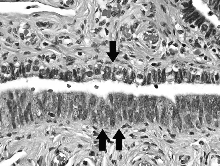
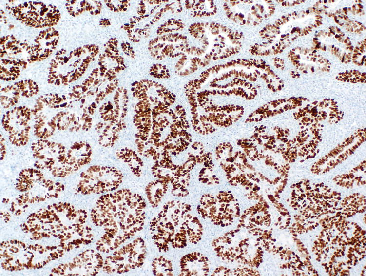
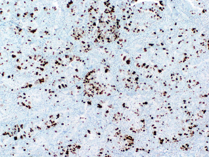
Ovarian serous carcinomas have been graded using various systems. Recently, a 2-tier system in which tumors are subdivided into low grade and high grade has been proposed. This approach is simplistic, reproducible, and based on biologic evidence indicating that both tumors develop via different pathways. Low-grade serous carcinomas exhibit low-grade nuclei with infrequent mitotic figures. They evolve from adenofibromas or borderline tumors, have frequent mutations of the KRAS, BRAF, or ERBB2 genes, and lack TP53 mutations (Type I pathway). The progression to invasive carcinoma is a slow step-wise process. Low-grade tumors are indolent and have better outcome than high-grade tumors. In contrast, high-grade serous carcinomas have high-grade nuclei and numerous mitotic figures. Identification of a precursor lesion in the ovary has been elusive and therefore the origin of ovarian carcinoma has been described as de novo. More recently, studies have suggested that a proportion seem to originate from intraepithelial carcinoma in the fallopian tube. The development of these tumors is rapid (Type II pathway). Most are characterized by TP53 mutations and lack mutations of KRAS, BRAF, or ERBB2. Although both types of serous carcinomas evolve along different pathways, rare high-grade serous carcinomas seem to arise through the Type I pathway. Immunohistochemical stains for p53, p16, and Ki-67 for distinction of low-grade from high-grade tumors are of limited value but can be helpful in selected instances. This review provides an update on the pathogenesis and clinicopathologic features of these 2 types of serous carcinomas and addresses some of the diagnostic problems that are encountered in routine practice.


Figures

Figure 1
Figure 1
Proposed model of pathogenesis of low-grade serous carcinoma. (A) Small atypical proliferative serous tumor (APST) arising in a serous cystadenoma. (B) APST. (C) Non-invasive micropapillary serous carcinoma (MPSC). (D) Tumor progression in the ovary results in invasive MPSC (lower); background of non-invasive MPSC in upper half of figure. (E) Microinvasion (upper left), which is qualitatively identical to low-grade serous carcinoma, arising within a non-invasive MPSC (lower right). (F) Invasive implants in the peritoneum are histologically identical to low-grade serous carcinoma.
Figure 1
Figure 1
Proposed model of pathogenesis of low-grade serous carcinoma. (A) Small atypical proliferative serous tumor (APST) arising in a serous cystadenoma. (B) APST. (C) Non-invasive micropapillary serous carcinoma (MPSC). (D) Tumor progression in the ovary results in invasive MPSC (lower); background of non-invasive MPSC in upper half of figure. (E) Microinvasion (upper left), which is qualitatively identical to low-grade serous carcinoma, arising within a non-invasive MPSC (lower right). (F) Invasive implants in the peritoneum are histologically identical to low-grade serous carcinoma.
Figure 2
Figure 2
Tubal intraepithelial carcinoma (TIC). (A) The epithelium of the fallopian tube mucosa with TIC is thicker compared with normal mucosa (upper center). (B) The neoplastic cells of TIC show enlarged nuclei, increased nuclear-to-cytoplasmic ratios, and coarse chromatin. (C) Immunohistochemical stain for p53 showing diffuse strong expression in TIC while the normal mucosa is negative. In (B) and (C), single arrow represents normal epithelium, and double arrows represent TIC.
Figure 2
Figure 2
Tubal intraepithelial carcinoma (TIC). (A) The epithelium of the fallopian tube mucosa with TIC is thicker compared with normal mucosa (upper center). (B) The neoplastic cells of TIC show enlarged nuclei, increased nuclear-to-cytoplasmic ratios, and coarse chromatin. (C) Immunohistochemical stain for p53 showing diffuse strong expression in TIC while the normal mucosa is negative. In (B) and (C), single arrow represents normal epithelium, and double arrows represent TIC.
Figure 3
Figure 3
High-grade serous carcinoma arising in a background of low-grade serous carcinoma. (A and B) Low-grade component. (C and D) High-grade component.
Figure 3
Figure 3
High-grade serous carcinoma arising in a background of low-grade serous carcinoma. (A and B) Low-grade component. (C and D) High-grade component.
Figure 3
Figure 3
High-grade serous carcinoma arising in a background of low-grade serous carcinoma. (A and B) Low-grade component. (C and D) High-grade component.
Figure 3
Figure 3
High-grade serous carcinoma arising in a background of low-grade serous carcinoma. (A and B) Low-grade component. (C and D) High-grade component.
Figure 4
Figure 4
Low-grade serous carcinoma. (A and B) Carcinoma invades stroma and is composed of small micropapillae. (B) The papillae either lack fibrous cores or contain thin attenuated fibrous cores and are surrounded by clear spaces. (C) In some instances, the papillae fuse to form anastamosing patterns. (D) Larger papillae may be seen. Psammoma bodies are also present. (E) The nuclei of the micropapillae are small, uniform, and round, with evenly dispersed chromatin and small nucleoli.
Figure 4
Figure 4
Low-grade serous carcinoma. (A and B) Carcinoma invades stroma and is composed of small micropapillae. (B) The papillae either lack fibrous cores or contain thin attenuated fibrous cores and are surrounded by clear spaces. (C) In some instances, the papillae fuse to form anastamosing patterns. (D) Larger papillae may be seen. Psammoma bodies are also present. (E) The nuclei of the micropapillae are small, uniform, and round, with evenly dispersed chromatin and small nucleoli.
Figure 5
Figure 5
Low-grade serous carcinoma with inverted macropapillary pattern. (A and B) The invasive papillae are medium to large in size, haphazardly arranged, and surrounded by clear spaces. The papillae are lined by cells with low-grade nuclei similar to those in the conventional micropapillary pattern.
Figure 5
Figure 5
Low-grade serous carcinoma with inverted macropapillary pattern. (A and B) The invasive papillae are medium to large in size, haphazardly arranged, and surrounded by clear spaces. The papillae are lined by cells with low-grade nuclei similar to those in the conventional micropapillary pattern.
Figure 6
Figure 6
High-grade serous carcinoma. (A) Typical papillary pattern showing irregular slit-like spaces. (B) Glandular pattern. (C) Diffuse, solid pattern. (D) Micropapillary pattern. (E) In contrast to low-grade serous carcinoma (Fig. 4E), the nuclei of high-grade serous carcinoma are larger with greater pleomorphism and larger nucleoli. (F) Endometrioid carcinoma-like pattern. (G) Serous carcinoma with clear cytoplasm. (H) Transitional cell carcinoma-like pattern. (I) Signet ring change simulating signet ring cells of metastatic adenocarcinoma involving the ovary.
Figure 6
Figure 6
High-grade serous carcinoma. (A) Typical papillary pattern showing irregular slit-like spaces. (B) Glandular pattern. (C) Diffuse, solid pattern. (D) Micropapillary pattern. (E) In contrast to low-grade serous carcinoma (Fig. 4E), the nuclei of high-grade serous carcinoma are larger with greater pleomorphism and larger nucleoli. (F) Endometrioid carcinoma-like pattern. (G) Serous carcinoma with clear cytoplasm. (H) Transitional cell carcinoma-like pattern. (I) Signet ring change simulating signet ring cells of metastatic adenocarcinoma involving the ovary.
Figure 7
Figure 7
High-grade serous carcinoma with “grade 2 nuclei.” (A and B) The nuclei are more uniform and smaller than in typical high-grade serous carcinomas but larger than low-grade serous carcinoma. The combination of (B) slightly increased variation in nuclear size, coarser chromatin, nucleolar prominence, and increased mitotic activity (arrows), (C) abnormal mitotic figures, and (D) necrosis allows for distinction from low-grade serous carcinoma.
Figure 7
Figure 7
High-grade serous carcinoma with “grade 2 nuclei.” (A and B) The nuclei are more uniform and smaller than in typical high-grade serous carcinomas but larger than low-grade serous carcinoma. The combination of (B) slightly increased variation in nuclear size, coarser chromatin, nucleolar prominence, and increased mitotic activity (arrows), (C) abnormal mitotic figures, and (D) necrosis allows for distinction from low-grade serous carcinoma.
Figure 8
Figure 8
Immunohistochemical staining for p53, p16, and Ki-67 in serous carcinoma. Low-grade carcinoma: (A) H&E, (B) p53 expression in scattered cells, (C) patchy p16 expression, and (D) low Ki-67 proliferation index. High-grade carcinoma: (E) H&E, (F) diffuse p53 expression, (G) diffuse p16 expression, and (H) high Ki-67 proliferation index. It should be noted that the H&E appearance in (E) is suggestive of a high-grade endometrioid carcinoma, but the immunophenotype is consistent with high-grade serous carcinoma.
Figure 8
Figure 8
Immunohistochemical staining for p53, p16, and Ki-67 in serous carcinoma. Low-grade carcinoma: (A) H&E, (B) p53 expression in scattered cells, (C) patchy p16 expression, and (D) low Ki-67 proliferation index. High-grade carcinoma: (E) H&E, (F) diffuse p53 expression, (G) diffuse p16 expression, and (H) high Ki-67 proliferation index. It should be noted that the H&E appearance in (E) is suggestive of a high-grade endometrioid carcinoma, but the immunophenotype is consistent with high-grade serous carcinoma.

References

    1. Silverberg SG. Histopathologic grading of ovarian carcinoma: a review and proposal. Int J Gynecol Pathol. 2000;19:7–15. - PubMed
    1. Shimizu Y, Kamoi S, Amada S, et al. Toward the development of a universal grading system for ovarian epithelial carcinoma: testing of a proposed system in a series of 461 patients with uniform treatment and follow-up. Cancer. 1998;82:893–901. - PubMed
    1. Ayhan A, Kurman RJ, Vang R, et al. Defining the cut-point between low- and high-grade ovarian serous carcinomas: a clinicopathologic and molecular genetic analysis. Am J Surg Pathol. (In Press) - PMC - PubMed
    1. Burks RT, Sherman ME, Kurman RJ. Micropapillary serous carcinoma of the ovary. A distinctive low-grade carcinoma related to serous borderline tumors. Am J Surg Pathol. 1996;20:1319–1330. - PubMed
    1. Dehari R, Kurman RJ, Logani S, et al. The development of high-grade serous carcinoma from atypical proliferative (borderline) serous tumors and low-grade micropapillary serous carcinoma: a morphologic and molecular genetic analysis. Am J Surg Pathol. 2007;31:1007–1012. - PubMed

Publication types

MeSH terms