

Genetic and phenotypic characteristics of pleomorphic lobular carcinoma in situ of the breast
- PMID: 19701073
- PMCID: PMC2783988
- DOI: 10.1097/PAS.0b013e3181b18a89
Genetic and phenotypic characteristics of pleomorphic lobular carcinoma in situ of the breast
Abstract
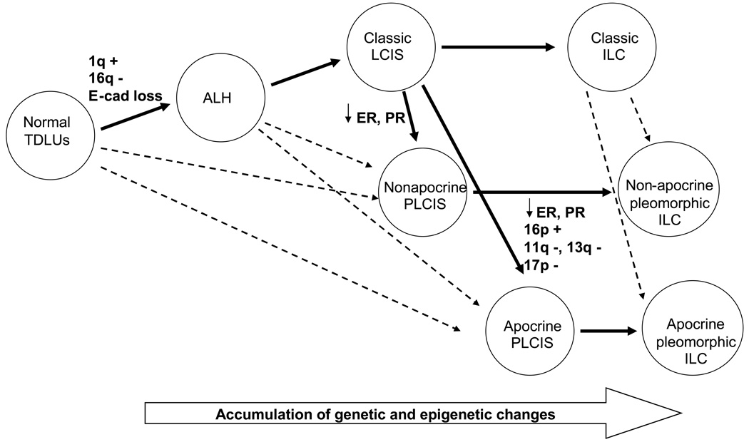
The clinical, pathologic, and molecular features of pleomorphic lobular carcinoma in situ (PLCIS) and the relationship of PLCIS to classic LCIS (CLCIS) are poorly defined. In this study, we analyzed 31 cases of PLCIS (13 apocrine and 18 nonapocrine subtypes) and compared the clinical, pathologic, immunophenotypic, and genetic characteristics of these cases with those of 24 cases of CLCIS. Biomarker expression was examined using immunostaining for E-cadherin, gross cystic disease fluid protein-15, estrogen, progesterone, androgen receptor, human epidermal growth factor receptor2, CK5/6, and Ki67. Array-based comparative genomic hybridization to assess the genomic alterations was performed using microdissected formalin-fixed paraffin-embedded samples. Patients with PLCIS presented with mammographic abnormalities. Histologically, the tumor cells were dyshesive and showed pleomorphic nuclei, and there was often associated necrosis and microcalcifications. All lesions were E-cadherin negative. Compared with CLCIS, PLCIS showed significantly higher Ki67 index, lower estrogen receptor and progesterone receptor expression, and higher incidence of HER2 gene amplification. The majority of PLCIS and CLCIS demonstrated loss of 16q and gain of 1q. Apocrine PLCIS had significantly more genomic alterations than CLCIS and nonapocrine PLCIS. Although lack of E-cadherin expression and the 16q loss and 1q gain-array-based comparative genomic hybridization pattern support a relationship to CLCIS, PLCIS has clinical, mammographic, histologic, immunophenotypic, and genetic features that distinguish it from CLCIS. The histologic features, biomarker profile, and genomic instability observed in PLCIS suggest a more aggressive phenotype than CLCIS. However, clinical follow-up studies will be required to define the natural history and most appropriate management of these lesions.
Figures


References
-
- Chivukula M, Haynik DM, Brufsky A, et al. Pleomorphic lobular carcinoma in situ (PLCIS) on breast core needle biopsies: clinical significance and immunoprofile. Am J Surg Pathol. 2008;32:1721–1726. - PubMed
-
- DeVries S, Gray JW, Pinkel D, et al. Comparative genomic hybridization Chapter 4. In: Haines Jonathan L, et al., editors. Current protocols in human genetics/editorial board. 2001. Unit46. - PubMed
-
- Ernster VL, Barclay J. Increases in ductal carcinoma in situ (DCIS) of the breast in relation to mammography: a dilemma. J Natl Cancer Inst Monogr. 1997;151(156) - PubMed
-
- Fadare O, Dadmanesh F, Alvarado-Cabrero I, et al. Lobular intraepithelial neoplasia [lobular carcinoma in situ] with comedo-type necrosis: A clinicopathologic study of 18 cases. Am J Surg Pathol. 2006;30:1445–1453. - PubMed
-
- Fisher ER, Costantino J, Fisher B, et al. Pathologic findings from the National Surgical Adjuvant Breast Project (NSABP) Protocol B-17. Five-year observations concerning lobular carcinoma in situ. Cancer. 1996;78:1403–1416. - PubMed
Publication types
MeSH terms
Substances
Grants and funding
LinkOut - more resources
Full Text Sources
Medical
Research Materials
Miscellaneous
