. 2009 Sep 10;461(7261):225-9.
doi: 10.1038/nature08265. Epub 2009 Aug 23.

Co-translational mRNA decay in Saccharomyces cerevisiae

Affiliations

Co-translational mRNA decay in Saccharomyces cerevisiae

Wenqian Hu et al. Nature. .

Abstract

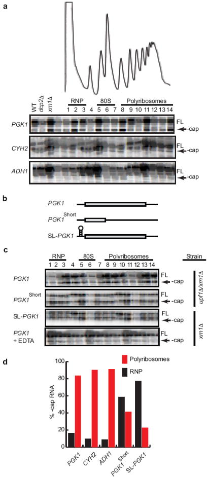
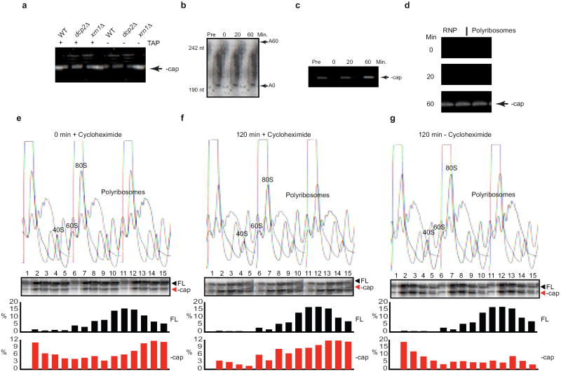
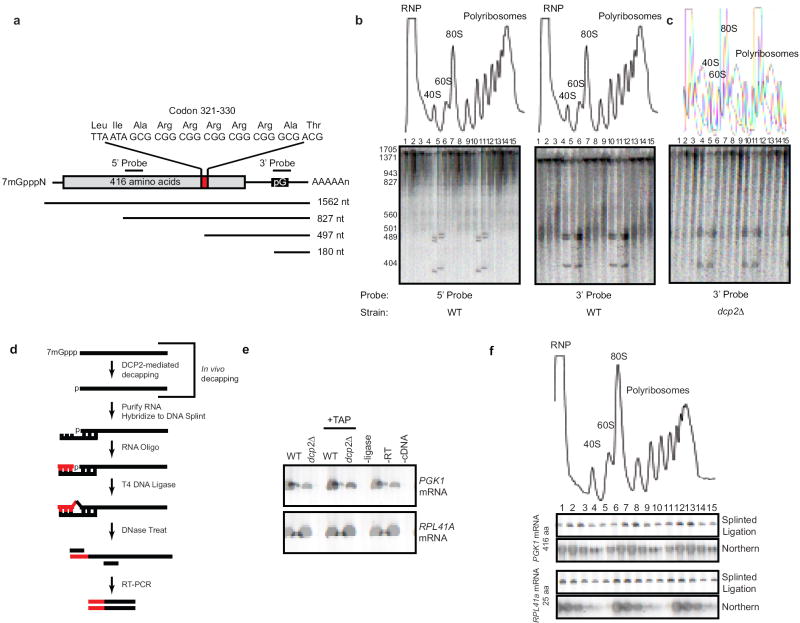
The rates of RNA decay and transcription determine the steady-state levels of all messenger RNA and both can be subject to regulation. Although the details of transcriptional regulation are becoming increasingly understood, the mechanism(s) controlling mRNA decay remain unclear. In yeast, a major pathway of mRNA decay begins with deadenylation followed by decapping and 5'-3' exonuclease digestion. Importantly, it is hypothesized that ribosomes must be removed from mRNA before transcripts are destroyed. Contrary to this prediction, here we show that decay takes place while mRNAs are associated with actively translating ribosomes. The data indicate that dissociation of ribosomes from mRNA is not a prerequisite for decay and we suggest that the 5'-3' polarity of mRNA degradation has evolved to ensure that the last translocating ribosome can complete translation.


Figures

Figure 1
Figure 1. Decapped mRNA is associated with polyribosomes
(a) Primer extension analysis on endogenous PGK1, CYH2, and ADH1 mRNA was performed on RNA isolated from sucrose gradient fractions of an xrn1Δ cell lysate. RNP, 80S, and polyribosomes are indicated above fraction numbers. FL, full length mRNA; - cap, decapped mRNA. Primer extension analyses on total RNA (15 μg) from WT, dcp2Δ, and xrn1Δ cells are shown on left side of each panel to indicate −cap mRNA is observed only in xrn1Δ cells. (b) Representation of PGK1 reporter, PGK1 reporter with a PTC (PGK1short), and PGK1 reporter with a stem-loop in its 5’ UTR (SL-PGK1). (c) Primer extension on RNA from sucrose gradient fractions from lysates of upf1Δ/xrn1Δ cells expressing PGK1 reporter or PGK1short reporter, and from xrn1Δ cells expressing SL-PGK1 or PGK1 reporter. In the bottom panel, lysates from xrn1Δ cells expressing the PGK1 reporter were incubated in presence of 50 mM EDTA prior to loading on sucrose gradients. (d) Quantification of – cap mRNAs as a percentage of total reverse transcription product in RNP and polyribosome fractions.
Figure 2
Figure 2. mRNA decapping is initiated on polyribosomes
All experiments in Fig. 2 were performed using cells expressing PGK1 reporter under control of the GAL1 promoter. (a) Total RNA from WT, dcp2Δ, and xrn1Δ cells was treated with (+) or without (-) tobacco acid pyrophosphatase (TAP) and circularization RT-PCR (cRT-PCR) was performed to detect decapped PGK1 reporter. (b-d) Transcriptional pulse-chase of PGK1 performed in xrn1Δ cells. (b) Poly(A) tail status of PGK1 was analyzed by oligonucleotide-directed RNase H cleavage, PAGE and Northern analysis. (c) Decapping of PGK1 mRNA monitored by cRT-PCR. (d) Cell lysates from the pulse-chase were separated on sucrose gradients. RNA from gradient fractions was pooled into non-translating (RNP) and polysome pools and decapped PGK1 was detected by cRT-PCR. (e-g) Transcriptional shut-off of PGK1 was performed in xrn1Δ cells. Lysates from cells at 0 min after shut-off (e), 120 min after shut-off in the presence of 25 μg/mL cycloheximide (f), and 120 min after shut-off without cycloheximide (g) were separated ny sucrose gradients. RNA from gradient fractions was analyzed by primer extension for PGK1 reporter. The quantifications of full length (FL) and decapped (-cap) mRNA as a percentage of total extension product are shown for each time point.
Figure 3
Figure 3. mRNA decapping occurs on polyribosomes in wild-type cells
The PGK1RC reporter is depicted (a). (b) Northern blot analysis of PGK1RC mRNA after sucrose gradient fractionation. RNA detected using a 5’ or 3’ probe as depicted in (a). The same analysis as in (b) performed in dcp2Δ cells (c). (d) Splinted-ligation RT-PCR assay to detect endogenous decapped mRNA in wild-type cells. An RNA adaptor is ligated specifically to decapped mRNA via a DNA splint by T4 DNA ligase. The DNA splint is removed by DNase I treatment and ligation product is detected by RT-PCR using a gene and adaptor specific primers. The PCR product is indicative of decapped mRNA. (e) Splinted-ligation RT-PCR analysis for endogenous PGK1 and RPL41A mRNAs on total RNA from WT and dcp2Δ cells. + TAP: total RNA treated with tobacco acid pyrophosphatase to remove the 5’ cap in vitro; −ligase: no T4 ligase; -RT: no reverse transcriptase; -cDNA: no cDNA template added to PCR. (f) RNA recovered from sucrose gradient fractions of wild-type cell lysate was analyzed by splinted-ligation RT-PCR or Northern blot using gene specific probe.

References

    1. Franks TM, Lykke-Andersen J. The control of mRNA decapping and P-body formation. Mol Cell. 2008;32(5):605–615. - PMC - PubMed
    1. Parker R, Sheth U. P bodies and the control of mRNA translation and degradation. Mol Cell. 2007;25(5):635–646. - PubMed
    1. Coller J, Parker R. Eukaryotic mRNA decapping. Annu Rev Biochem. 2004;73:861–890. - PubMed
    1. Eulalio A, Behm-Ansmant I, Izaurralde E. P bodies: at the crossroads of post-transcriptional pathways. Nat Rev Mol Cell Biol. 2007;8(1):9–22. - PubMed
    1. He F, Jacobson A. Upf1p, Nmd2p, and Upf3p regulate the decapping and exonucleolytic degradation of both nonsense-containing mRNAs and wild-type mRNAs. Mol Cell Biol. 2001;21(5):1515–1530. - PMC - PubMed

Publication types