

Activation of innate immune antiviral responses by Nod2
- PMID: 19701189
- PMCID: PMC2752345
- DOI: 10.1038/ni.1782
Activation of innate immune antiviral responses by Nod2
Erratum in
- Nat Immunol. 2010 Oct;11(10):969
Abstract
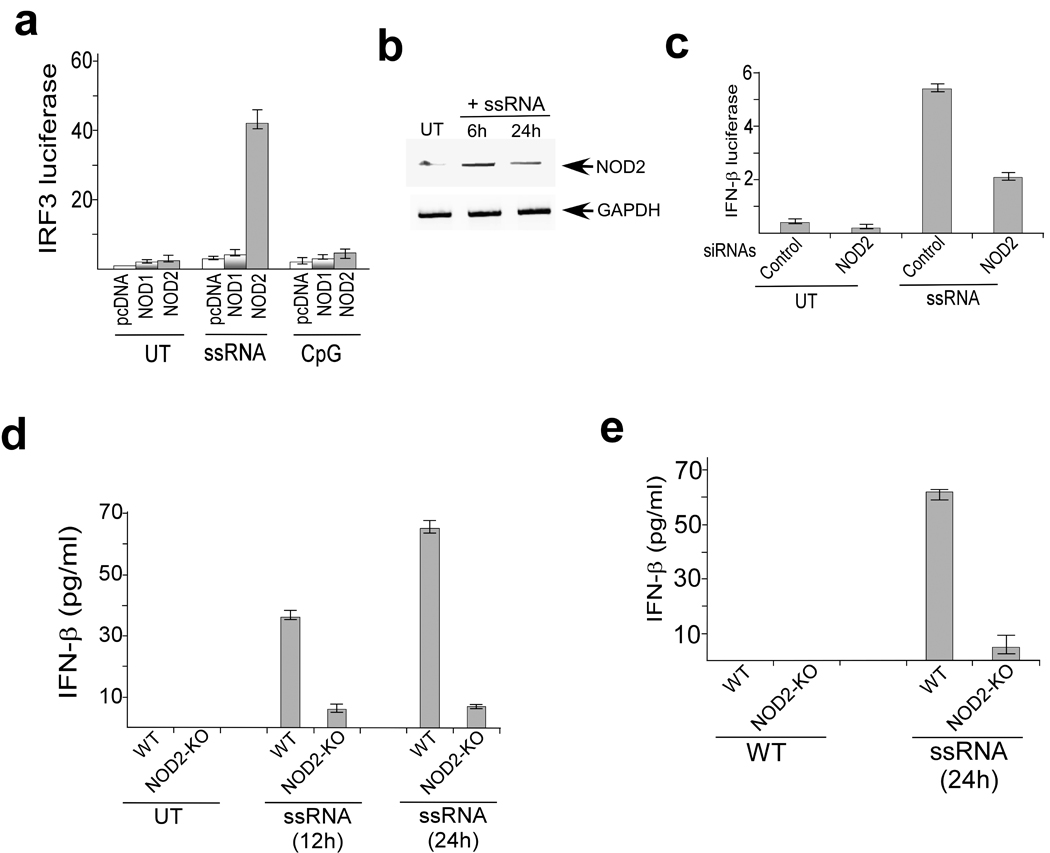
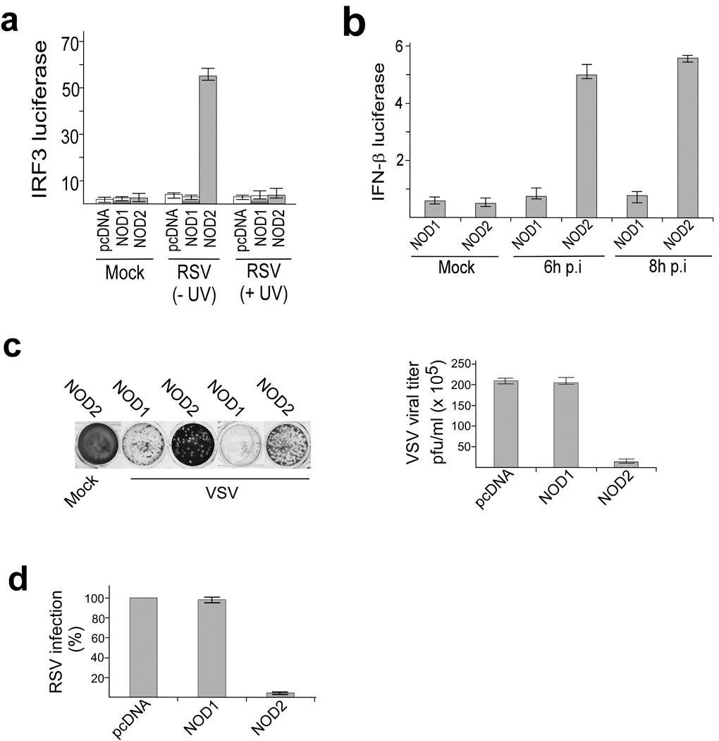
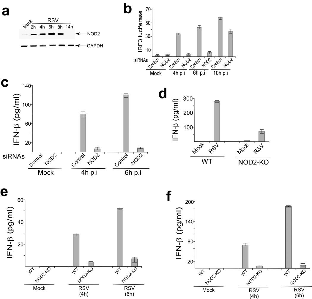
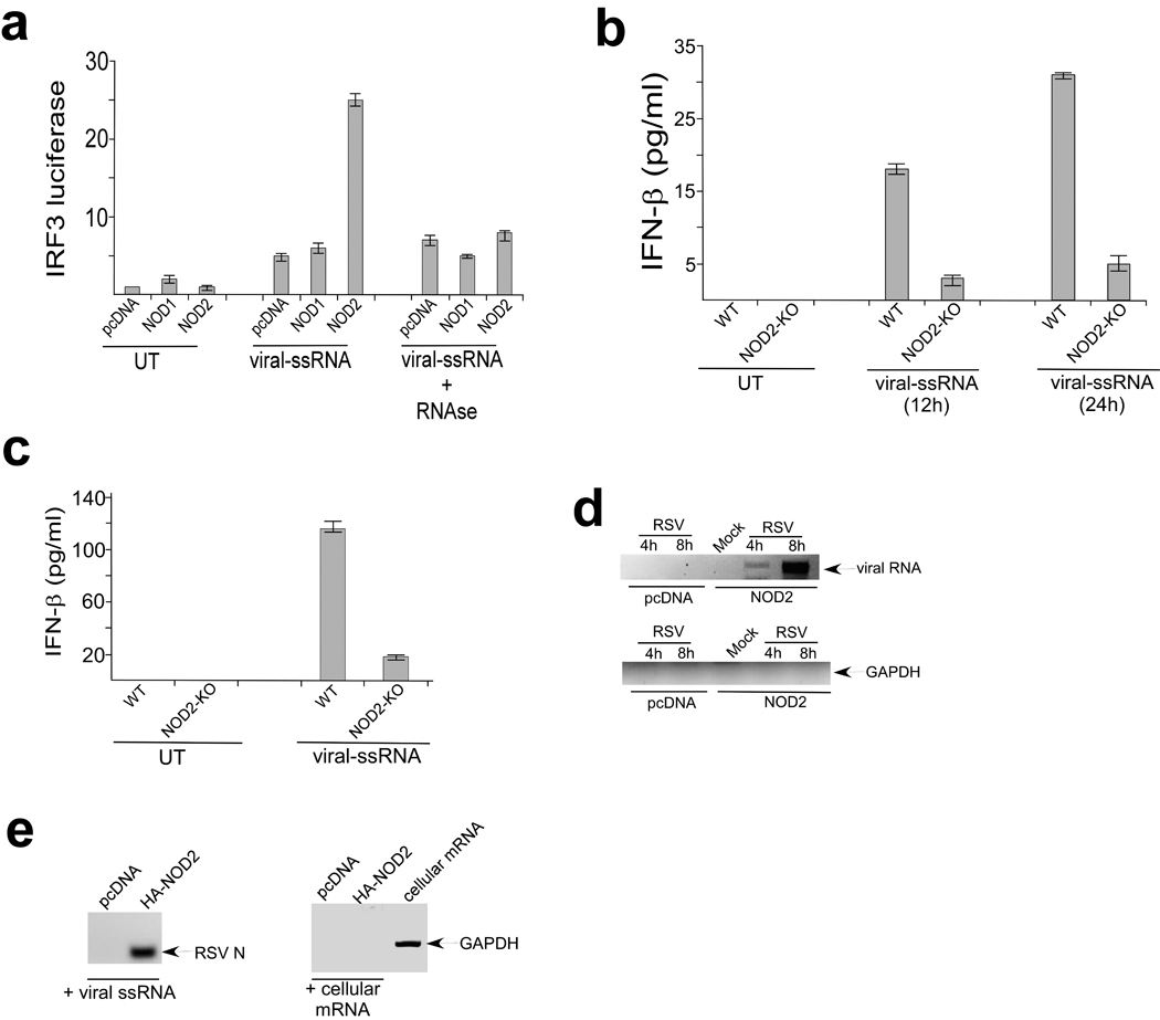
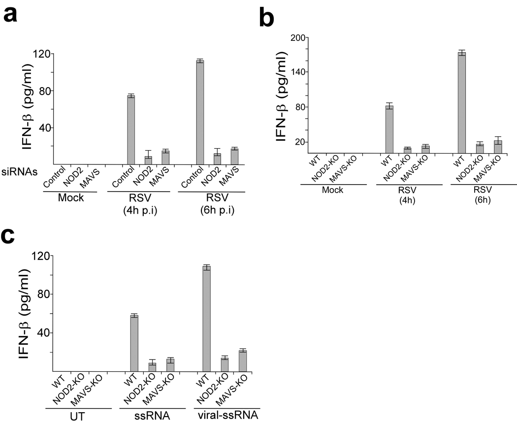
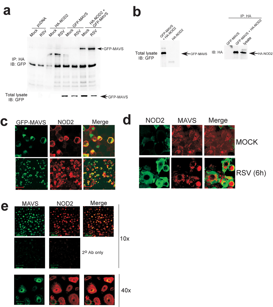
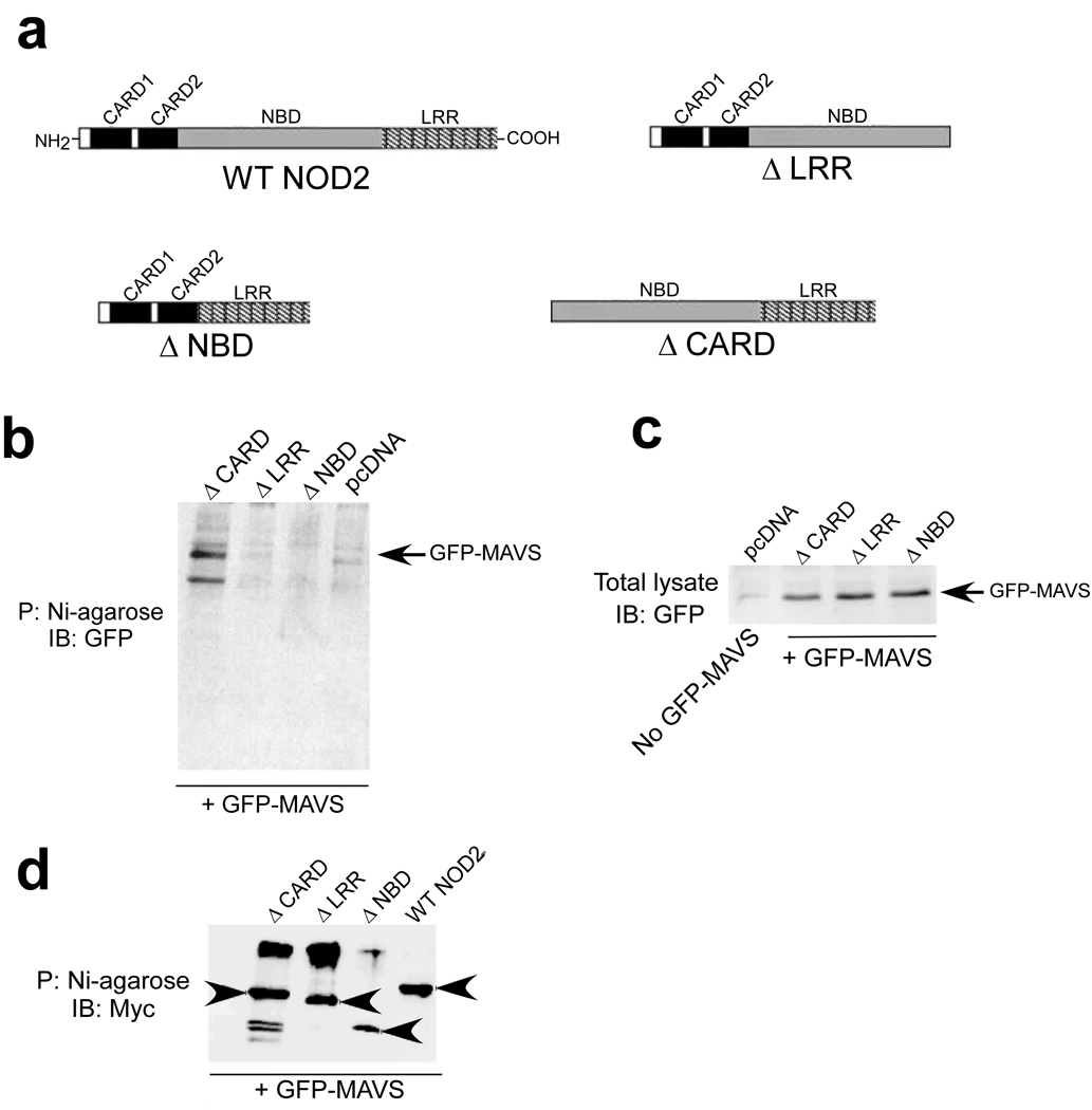
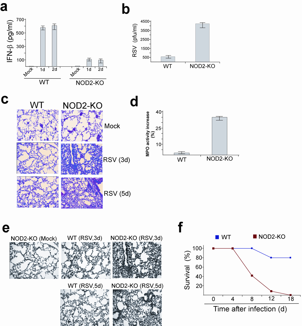
Pattern-recognition receptors (PRRs), including Toll-like receptors (TLRs) and RIG-like helicase (RLH) receptors, are involved in innate immune antiviral responses. Here we show that nucleotide-binding oligomerization domain 2 (Nod2) can also function as a cytoplasmic viral PRR by triggering activation of interferon-regulatory factor 3 (IRF3) and production of interferon-beta (IFN-beta). After recognition of a viral ssRNA genome, Nod2 used the adaptor protein MAVS to activate IRF3. Nod2-deficient mice failed to produce interferon efficiently and showed enhanced susceptibility to virus-induced pathogenesis. Thus, the function of Nod2 as a viral PRR highlights the important function of Nod2 in host antiviral defense mechanisms.
Figures
Comment in
-
Beyond peptidoglycan for Nod2.Nat Immunol. 2009 Oct;10(10):1053-4. doi: 10.1038/ni1009-1053. Nat Immunol. 2009. PMID: 19767725 No abstract available.
Similar articles
-
Leukotriene B4 Enhances NOD2-Dependent Innate Response against Influenza Virus Infection.PLoS One. 2015 Oct 7;10(10):e0139856. doi: 10.1371/journal.pone.0139856. eCollection 2015. PLoS One. 2015. PMID: 26444420 Free PMC article.
-
Nucleotide oligomerization and binding domain 2-dependent dendritic cell activation is necessary for innate immunity and optimal CD8+ T Cell responses to influenza A virus infection.J Virol. 2014 Aug;88(16):8946-55. doi: 10.1128/JVI.01110-14. Epub 2014 May 28. J Virol. 2014. PMID: 24872587 Free PMC article.
-
Phosphatidylinositol-3-kinase and Akt are required for RIG-I-mediated anti-viral signalling through cross-talk with IPS-1.Immunology. 2015 Feb;144(2):312-20. doi: 10.1111/imm.12373. Immunology. 2015. PMID: 25158146 Free PMC article.
-
Regulation of antiviral innate immune signaling by stress-induced RNA granules.J Biochem. 2016 Mar;159(3):279-86. doi: 10.1093/jb/mvv122. Epub 2016 Jan 8. J Biochem. 2016. PMID: 26748340 Free PMC article. Review.
-
The Role of Nucleotide-Binding Oligomerization Domain-Like Receptors in Pulmonary Infection.Am J Respir Cell Mol Biol. 2017 Aug;57(2):151-161. doi: 10.1165/rcmb.2016-0375TR. Am J Respir Cell Mol Biol. 2017. PMID: 28157451 Free PMC article. Review.
Cited by
-
RNA helicase signaling is critical for type i interferon production and protection against Rift Valley fever virus during mucosal challenge.J Virol. 2013 May;87(9):4846-60. doi: 10.1128/JVI.01997-12. Epub 2013 Feb 13. J Virol. 2013. PMID: 23408632 Free PMC article.
-
Newly described pattern recognition receptors team up against intracellular pathogens.Nat Rev Immunol. 2013 Aug;13(8):551-65. doi: 10.1038/nri3479. Epub 2013 Jul 12. Nat Rev Immunol. 2013. PMID: 23846113 Review.
-
Enter at your own risk: how enteroviruses navigate the dangerous world of pattern recognition receptor signaling.Cytokine. 2013 Sep;63(3):230-6. doi: 10.1016/j.cyto.2013.05.007. Epub 2013 Jun 10. Cytokine. 2013. PMID: 23764548 Free PMC article. Review.
-
Emerging role of ubiquitination in antiviral RIG-I signaling.Microbiol Mol Biol Rev. 2012 Mar;76(1):33-45. doi: 10.1128/MMBR.05012-11. Microbiol Mol Biol Rev. 2012. PMID: 22390971 Free PMC article. Review.
-
Highlights of the advances in basic immunology in 2011.Cell Mol Immunol. 2012 May;9(3):197-207. doi: 10.1038/cmi.2012.12. Epub 2012 Apr 23. Cell Mol Immunol. 2012. PMID: 22522654 Free PMC article. Review.
References
-
- Kawai T, Akira S. Innate immune recognition of viral infection. Nat. Immunol. 2006;7:131–137. - PubMed
-
- Bose S, Banerjee AK. Innate immune response against nonsegmented negative strand RNA viruses. J. Interferon. Cytokine. Res. 2003;23:401–412. - PubMed
-
- Stark GR, Kerr IM, Williams BRG, Silverman RH, Schreiber RD. How cells respond to interferons. Annu. Rev. Biochem. 1998;67:227–264. - PubMed
-
- Uematsu S, Akira S. Toll-like receptors and Type I interferons. J. Biol. Chem. 2007;282:15319–15323. - PubMed
-
- O'Neill LA. How Toll-like receptors signal: what we know and what we don't know. Curr. Opin. Immunol. 2006;18:3–9. - PubMed
Publication types
MeSH terms
Substances
Grants and funding
- R21 CA129246/CA/NCI NIH HHS/United States
- P01AG19316/AG/NIA NIH HHS/United States
- CA129246/CA/NCI NIH HHS/United States
- P01 AG019316/AG/NIA NIH HHS/United States
- AI069062/AI/NIAID NIH HHS/United States
- AI067716/AI/NIAID NIH HHS/United States
- R21 AI069062/AI/NIAID NIH HHS/United States
- P30 CA054174/CA/NCI NIH HHS/United States
- R01 AI067716/AI/NIAID NIH HHS/United States
- T32 DE014318/DE/NIDCR NIH HHS/United States
- P30 CA54174/CA/NCI NIH HHS/United States
- T32-DE14318/DE/NIDCR NIH HHS/United States
- P30 AG013319/AG/NIA NIH HHS/United States
LinkOut - more resources
Full Text Sources
Other Literature Sources
Molecular Biology Databases
Miscellaneous
