. 2009 Sep;41(9):977-85.
doi: 10.1038/ng.435. Epub 2009 Aug 23.

Posterior malformations in Dact1 mutant mice arise through misregulated Vangl2 at the primitive streak

Affiliations

Posterior malformations in Dact1 mutant mice arise through misregulated Vangl2 at the primitive streak

Rowena Suriben et al. Nat Genet. 2009 Sep.

Abstract

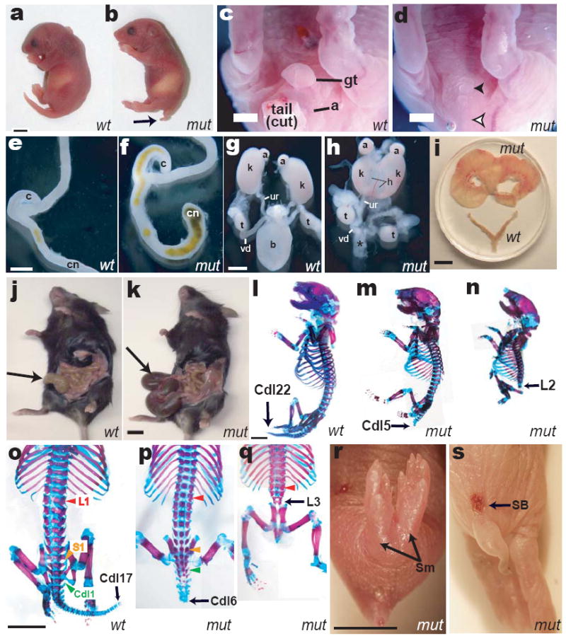
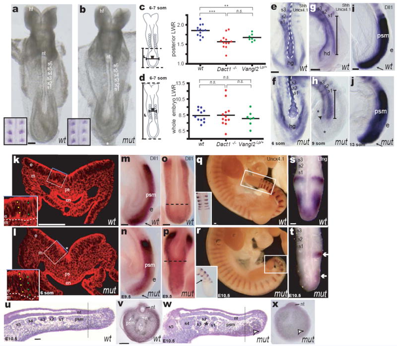
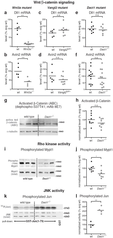
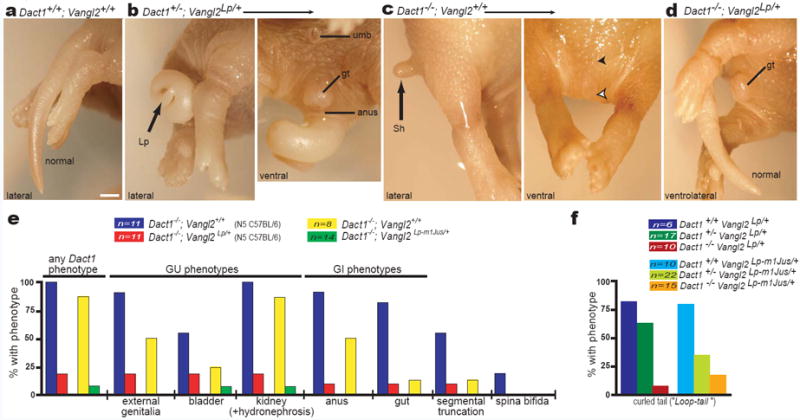
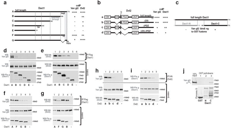
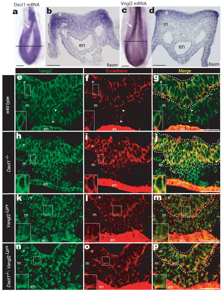
Mice homozygous for mutations in Dact1 (also called Dapper or Frodo) phenocopy human malformations involving the spine, genitourinary system and distal digestive tract. We traced this phenotype to disrupted germ-layer morphogenesis at the primitive streak. Notably, heterozygous mutation of Vangl2, a transmembrane component of the planar cell polarity (PCP) pathway, rescued recessive Dact1 phenotypes, whereas loss of Dact1 reciprocally rescued semidominant Vangl2 phenotypes. We show that Dact1, an intracellular protein, forms a complex with Vangl2. In Dact1 mutants, Vangl2 was increased at the primitive streak, where cells ordinarily undergo an epithelial-mesenchymal transition. This is associated with abnormal E-cadherin distribution and changes in biochemical measures of the PCP pathway. We conclude that Dact1 contributes to morphogenesis at the primitive streak by regulating Vangl2 upstream of cell adhesion and the PCP pathway.


Figures

Figure 1
Figure 1
Birth phenotypes in Dact1 mutants (mut) compared to wild type (wt). a, b Outward appearance; arrow, short tail. c, d Genital tubercle (gt) and anus (a) are missing in mutants (filled arrowhead, empty arrowhead), along with the tail. e, f Mutants have a blind-ended colon (cn). g, h Mutants lack bladder (b vs. *), have malformed hydronephrotic (h) kidneys (k, fused in the mutant specimen), and misconnected ureters (ur, connected to the vas deferens (vd) in this mutant male). i-k, Phenotypes of rare surviving adult Dact1 mutants consistent with impaired uterine outflow resulting in hydrometrocolpos (top vs. bottom in i), and impaired digestive tract evacuation resulting in megacolon (arrow in mutant k vs. wild type j). l-q Skeletons; black arrows indicate identity of the terminal ossified vertebra, colored arrowheads in o-q indicate segmental levels: red, lumbar-1 (L1); yellow, sacral-1 (S1); green, caudal-1 (Cdl1). r Sirenomelia (Sm). s Spina bifida (SB). Other abbreviations: (a) adrenal, (c) cecum, (t) testis. Scale bars: 0.5 mm, except i-k 5 mm.
Figure 2
Figure 2
Dact1 mutant embryonic phenotypes. a, b Early Dact1 mutant (mut) embryos appear normal except for their posterior contour (*); insets: Uncx4.1 WISH (somites). c, d Length-Width-Ratio (LWR) measurements in wild type (blue), Dact1 mutants (red) and Vangl2Lp heterozygotes (green) posterior (c) or whole (d) embryo. e-h Shh/Uncx4.1 WISH. e, f ventral aspect; g, h lateral aspect. Notochord (outline in e, f; bracket in g, h) is shorter and broader in Dact1 mutants compared to wild type. Hindgut diverticulum (hd in g) has not formed in mutant (arrowhead in h). Mesenchymal tissue (*) surrounds foreshortened axial structures. i, j Dll1 WISH: presomitic mesoderm (psm) and ectoderm (e, arrow) length are normal. k, l Phalloidin-stained transverse section at the primitive streak (ps). In mutant (l) endoderm (en) has not closed ventrally and is thinner, whereas ectoderm (e) is thicker (insets) and more sharply folded (*). m-p At later stages, Dll WISH (m, n lateral aspect; o, p dorsal aspect) reveals presomitic mesoderm and ectoderm of normal length (arrows), but less extended axial tissues (dotted lines). q, r Segmental abnormalities confined to the newest somites in the tail bud (insets) can be detected by Uncx4.1 WISH at later stages (insets: close-up and dorsal view). s, t Somitogenic clock abnormalities in Dact1 mutant tail buds revealed by abnormal asymmetric Lfng WISH (arrows). u-x Representative parasagittal (u, w); and transverse (v, x) sections through wild type (u, v) and Dact1 mutant (w, x) tail buds at E10.5. Dact1 mutants with abnormal somites (asterisk in w) also have severely disrupted posterior tissues and pooled blood at their ventral posterior tip (arrowhead in w, x); the neural tube (nt) nonetheless extends toward the tail bud tip. Dotted lines in u, w indicate approximate position of transverse sections from different embryos in v, x. Other abbreviations: (hf) head-folds, (s1-s8) somites, (hg) hindgut, (m) mesoderm, (n) notochord, (tg) tail gut. Scale bars = 0.1 mm. Statistical analysis (c, d): parametric unpaired two-tailed t test, horizontal line = mean; n.s. p > 0.05, **p < 0.01, ***p < 0.001
Figure 3
Figure 3
Biochemical correlates of PCP signaling are specifically affected at early phenotypic stages. Insets: assays were conducted on posterior tissue lysates from E8 and E9 embryos as shown. All graphs: closed circles, wild type (+/+); open circles, mutant littermates. a-h Wnt/β-catenin assays. QPCR for endogenous targets of Wnt/β-catenin signaling at E8 reveals that Dll1 and Axin2 mRNA levels are significantly reduced in Wnt3a null embryos (a, b), but not in Vangl2Lp heterozygotes (c, d), nor in Dact1 mutants (e, f). Similarly, levels of unphosphorylated (activated) β-catenin protein are not significantly reduced in Dact1 mutants at E9 (g, h). i-l PCP pathway biochemistry. Phosphorylation of Mypt1, a target of Rho kinase, is significantly reduced in Dact1 mutants at E8 (i, j), whereas JNK activity is robustly increased at E9 (k, l). Statistical analyses: parametric unpaired two-tailed t tests, horizontal line = mean; n.s. p > 0.05, *p < 0.05, ***p < 0.001
Figure 4
Figure 4
Mutations in Dact1 and Vangl2 mutually rescue. a-d Posterior phenotypes of littermates from a Dact1 × Vangl2 mutant intercross. a Dact1+/+;Vangl2+/+ neonate (wild type) has a normal tail. b Dact1+/-;Vangl2Lp/+ neonate (trans-heterozygous) has a curly tail (Lp, left panel) typical of the semidominant Loop-tail phenotype, while the genital tubercle (gt) and anus (a) are normal (as expected for a Dact1 heterozygote, right panel). c Dact1-/-;Vangl2+/+ neonate has the shortened tail (Sh; left panel), absent genital tubercle and anus (filled and empty arrowheads, right panel) typical of Dact1 mutants. d Dact1-/-;Vangl2Lp/+ combination mutant neonate (genetically rescued mutant) has a normal genital tubercle (gt), anus (not shown), and tail. e Quantitation of phenotypes and affected organs in Dact1 mutant littermates that either carry (red) or don’t carry (blue) the Vangl2Lp allele, or that either carry (green) or don’t carry (yellow) the Vangl2Lp-m1Jus allele (see also Supplementary Table 3a). f The Dact1 mutant allele reciprocally rescues, in a dose-dependent manner, Loop-tail phenotypes produced by heterozygosity for either Vangl2Lp (left, darker bars) or Vangl2Lp-m1Jus (right, lighter bars) (see also Supplementary Table 3b). Other abbreviations: (umb) umbilicus. Scale bar: 1 mm.
Figure 5
Figure 5
The Dact1 and Vangl2 proteins associate independently of Dvl. a-c Schematic summaries of coIP and GST pull-down data. d-i coIPs of proteins recombinantly expressed in human embryonic kidney cells. d Vangl2 coIPs with full-length Dact1, but loses affinity as the Dact1 C-terminus is progressively deleted. e Dvl2 coIPs with Dact1 unless both the C-terminus and middle region are deleted. f, g Separate C-terminal subregions of Dact1 mediate coIP with Vangl2 (f) versus Dvl2 (g). h Vangl2 coIPs with full-length Dvl2 unless the Dvl2 PDZ domain is deleted; there is also a slight contribution from the Dvl2 DS domain. i Dact1 coIPs with Dvl2 even in the absence of both these domains. j A GST-fusion protein incorporating the Dact1 C-terminus binds to Vangl2 protein synthesized in a wheat germ extract with no Dvl proteins.
Figure 6
Figure 6
Dact1 and Vangl2 functionally interact at the PS. a, b WISH Dact1 c, d WISH Vangl2 a, c Dorsal aspect showing neural fold (arrow) and approximate level of sections in b & d (dotted lines) b, d Transverse section at PS level. e-p Confocal fluorescent immunohistological localization of Vangl2 (green) and E-cadherin proteins (red) at the PS in Dact1+/+;Vangl2+/+ (wild type; e-g), Dact1-/-;Vangl2+/+ (Dact1 mutant; h-j), Dact1+/+;Vangl2Lp/+ (Loop-tail; k-m), and Dact1-/-;Vangl2Lp/+ (genetically rescued mutant; n-p). Abbreviations as in Figure 2. Scale bars in a-d 0.1 mm, in e-p 0.05mm.

Similar articles

Cited by

References

    1. Yoshikawa Y, Fujimori T, McMahon AP, Takada S. Evidence that absence of Wnt-3a signaling promotes neuralization instead of paraxial mesoderm development in the mouse. Dev Biol. 1997;183:234–42. - PubMed
    1. Torban E, et al. Genetic interaction between members of the Vangl family causes neural tube defects in mice. Proc Natl Acad Sci U S A. 2008;105:3449–54. - PMC - PubMed
    1. Wang J, et al. Dishevelled genes mediate a conserved mammalian PCP pathway to regulate convergent extension during neurulation. Development. 2006;133:1767–78. - PMC - PubMed
    1. Ybot-Gonzalez P, et al. Convergent extension, planar-cell-polarity signalling and initiation of mouse neural tube closure. Development. 2007;134:789–99. - PMC - PubMed
    1. Veeman MT, Axelrod JD, Moon RT. A second canon: Functions and mechanisms of beta-catenin-independent wnt signaling. Developmental Cell. 2003;5:367–377. - PubMed

Publication types

MeSH terms

Substances