. 2009 Oct;15(10):1208-14.
doi: 10.1038/nm.2019. Epub 2009 Aug 23.

Arrested maturation of excitatory synapses in autosomal dominant lateral temporal lobe epilepsy

Affiliations

Arrested maturation of excitatory synapses in autosomal dominant lateral temporal lobe epilepsy

Yu-Dong Zhou et al. Nat Med. 2009 Oct.

Abstract

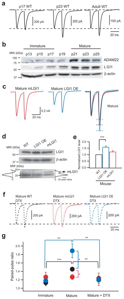
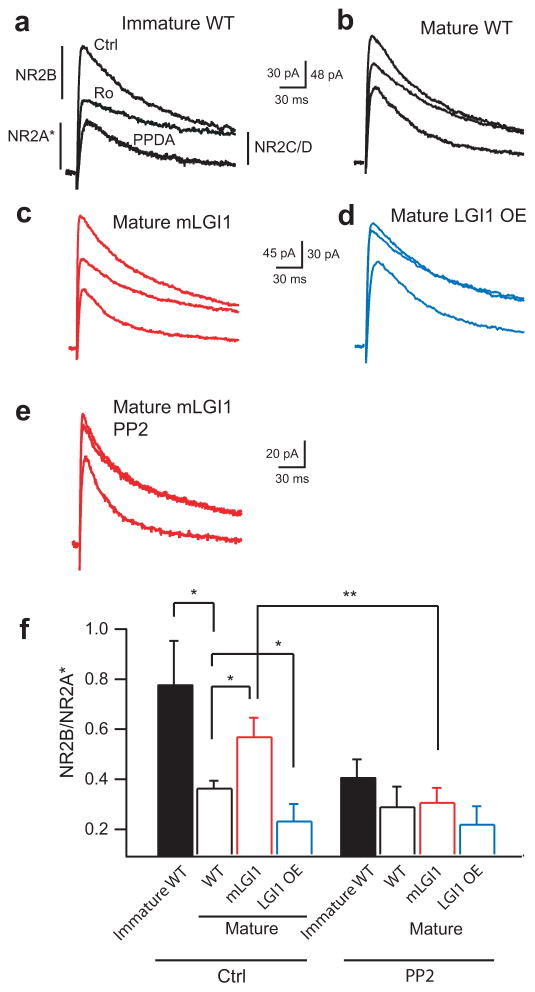
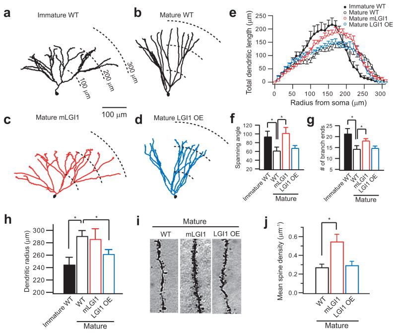
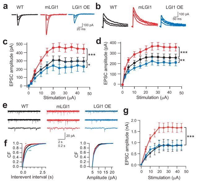
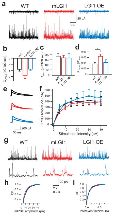
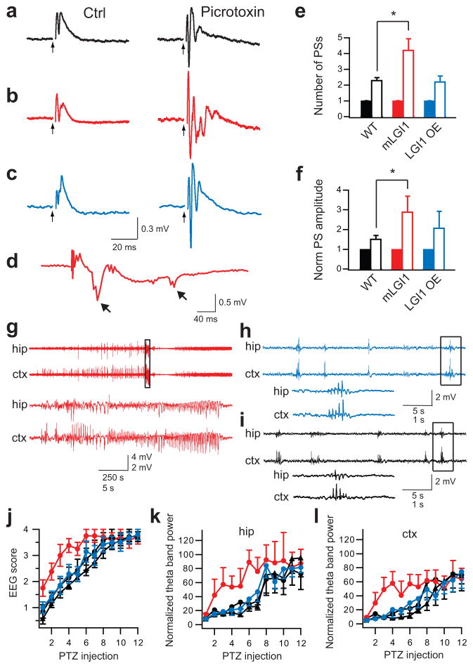
A subset of central glutamatergic synapses are coordinately pruned and matured by unresolved mechanisms during postnatal development. We report that the human epilepsy gene LGI1, encoding leucine-rich, glioma-inactivated protein-1 and mutated in autosomal dominant lateral temporal lobe epilepsy (ADLTE), mediates this process in hippocampus. We created transgenic mice either expressing a truncated mutant LGI1 (835delC) found in ADLTE or overexpressing a wild-type LGI1. We discovered that the normal postnatal maturation of presynaptic and postsynaptic functions was arrested by the 835delC mutant LGI1, and contrastingly, was magnified by excess wild-type LGI1. Concurrently, mutant LGI1 inhibited dendritic pruning and increased the spine density to markedly increase excitatory synaptic transmission. Inhibitory transmission, by contrast, was unaffected. Furthermore, mutant LGI1 promoted epileptiform discharge in vitro and kindling epileptogenesis in vivo with partial gamma-aminobutyric acid(A) (GABA(A)) receptor blockade. Thus, LGI1 represents a human gene mutated to promote epilepsy through impaired postnatal development of glutamatergic circuits.


Figures

Figure 1
Figure 1
LGI1 down-regulates presynaptic release probability at hippocampal MPP-GC excitatory synapses via Kv1 channels during postnatal development. (a) Representative traces showing paired-pulse facilitation (PPF) of evoked MPP-GC postsynaptic currents, a measure of release probability, increases between postnatal days 17 and 23 in control (WT) mice. (b) Western blots showing LGI1 and its receptor, ADAM22, also increase between p17 and p23. (c) Representative traces showing PPF remains low in mature mLGI1 and significantly increases in mature LGI1 OE mice relative to control. (d) Western blot showing increased expression of 64 kDa LGI1 in LGI1 OE mice and expression of 23 kDa mutant LGI1, marked by an arrow on densitometry traces, in mLGI1 mice. (e) Quantification of 64 kDa LGI1 protein density (normalized to β-actin) in WT, LGI1 OE, and mLGI1 mice (n = 7). (f) Kv1 channel blocker α-dendrotoxin (DTX) eliminates the difference in paired-pulse ratio (ppr) in mature mice. (g) Quantification of ppr in WT (black symbols), mLGI1 (red symbols), and LGI1 OE (blue symbols) mice (n = 4 to 25). *: P < 0.05; **: P < 0.01; ***: P < 0.001.
Figure 2
Figure 2
LGI1 down-regulates postsynaptic NR2B-dependent NMDA receptor currents during postnatal brain development. (a,b) Representative current traces from wild-type (WT) showing NR2B subunit-dependent current is larger in immature (a) than mature (b) wild-type GCs. Ro: NR2B blocker; PPDA: NR2C/2D blocker. (c,d) Representative current traces showing NR2B-dependent currents remain high in mature mLGI1 (c) and down-regulate excessively in mature LGI1 OE (d) transgenic mice relative to mature wild-type (b). (e) Representative traces showing Src kinase inhibitor, PP2, inhibits the NR2B component in mature mLGI1. (f) Quantification of NR2B/NR2A* current ratio in the three genotypes (n = 5 to 11) across postnatal development in the presence and absence of PP2. *: P < 0.05; **: P < 0.01.
Figure 3
Figure 3
ADLTE mutant LGI1 blocks dendritic pruning during postnatal development and increases spine density. (a,b) Representative Neurolucida-reconstructed wild-type (WT) GC dendrites showing a widely branched arbor in immature GCs (a) and a less branched arbor in mature GCs (b). (c,d) Representative Neurolucida-reconstructed GC dendrites showing a widely branched arbor in mature mLGI1 GCs (c) and a shortened arbor in mature LGI1 OE GCs (d). (e) Sholl analysis of GC dendritic arbor. (f–h) Quantification of dendritic spanning angle (f), branch end number (g), and mean radius (h) of immature WT (n = 7), mature WT (n = 6), mature mLGI1 (n = 7), and mature LGI1 OE (n = 7) GCs. (i,j) Representative images (i) and quantification of mean spine density(j) showing an increased spine number in mature mLGI1 GCs (n = 4) compared to mature WT (n = 5) and mature LGI1 OE (n = 6) GCs. *: P < 0.05.
Figure 4
Figure 4
ADLTE mutant LGI1 increases glutamatergic synaptic transmission. (a,b) Representative traces showing evoked AMPA (a) and NMDA (b) receptor-mediated EPSCs (2 μA, 5 μA, and saturating stimulus intensities) in wild-type (WT), mLGI1, and LGI1 OE mice. (c,d) Quantification of EPSC amplitude to stimulus intensity for AMPA (c, WT, black, n = 10; mLGI1, red, n = 7; LGI1 OE, blue, n = 6) and NMDA (d, WT, black, n = 11; mLGI1, red, n = 11; LGI1 OE, blue, n = 7) receptor currents. (e) Representative traces showing mEPSCs recorded from GCs of WT, mLGI1, and LGI1 OE transgenic mice. (f) Cumulative frequency plots of mEPSC interevent interval and amplitude for WT (black), mLGI1 (red), and LGI1 OE (blue) transgenic mice (n = 5). (g) Quantification of maximum evoked EPSC in DTX in WT (black symbol, n = 3), mLGI1 (red symbol, n = 4), and LGI1 OE (blue symbol, n = 3) mice. *:P < 0.05; **: P < 0.01; ***: P < 0.001.
Figure 5
Figure 5
LGI1 trangenes fail to effect inhibitory synaptic transmission. (a) Representative traces showing spontaneous EPSC (sEPSC, upper) and IPSC (sIPSC, lower) recorded in the same cell at –60 mV (upper) and +10 mV (lower) from wild-type (WT, black), mLGI1 (red), and LGI1 OE (blue) transgenic mice. (b–d) Quantification of sEPSC (b) and sIPSC (c) charge transfer and sEPSC/sIPSC charge transfer ratios (d) in WT (n = 10), mLGI1 (n = 9), and LGI1 OE (n = 4) mice. (e) Representative traces showing evoked GABAA receptor-mediated IPSC (5 μA, and saturating stimulus intensities) in WT (black), mLGI1 (red), and LGI1 OE (blue) mice. (f) Quantification of evoked IPSC amplitudes for WT (black), mLGI1 (red), and LGI1 OE (blue) mice (n = 4). (g) Representative traces showing miniature IPSC (mIPSC) recorded from GCs of wild-type, mLGI1, and LGI1 OE mouse. (h,i) Cumulative frequency plots of mIPSC amplitude (h) and interevent interval (i) for WT (black), mLGI1 (red), and LGI1 OE (blue) (n = 5). *: P < 0.05.
Figure 6
Figure 6
ADLTE mutant LGI1 promotes epilepsy. (a–c) Representative traces showing field potentials from the GC layer of wild-type (WT) (a), mLGI1 (b), and LGI1 OE (c) mice in response to MPP stimulation (arrow heads) before (left) and after (right) picrotoxin (PTX) treatment. (d) Representative trace showing afterdischarges (arrows) followed population spikes only in PTX-treated mLGI1 slices. (e,f) Quantification of mean population spike (PS) number (e) and normalized maximum amplitude (f) for WT (black) (n = 7), mLGI1 (red) (n = 5), and LGI1 OE (blue) (n = 6) mice before (solid bars) and after (open bars) PTX treatment. (g–i) Representative EEG traces from mLGI1 (g), LGI1 OE (h), and WT littermate (i) mice after 6th PTZ injection. The areas marked by the rectangles are expanded in the lower traces. (j–l) Quantifications of EEG scores (j), normalized hippocampal (k), and cortical (l) EEG theta band power of PTZ-kindling in mLGI1 mice (red circles) (n = 8) vs. their wild-type littermates (black circles) (n = 8), and LGI1 OE mice (blue circle) (n = 7) vs. their wild-type littermates (black triangles) (n = 7). *: P < 0.05.

Comment in

References

    1. Waites CL, Craig AM, Garner CC. Mechanisms of vertebrate synaptogenesis. Annu Rev Neurosci. 2005;28:251–274. - PubMed
    1. Chen C, Regehr WG. Developmental remodeling of the retinogeniculate synapse. Neuron. 2000;28:955–966. - PubMed
    1. Rihn LL, Claiborne BJ. Dendritic growth and regression in rat dentate granule cells during late postnatal development. Brain Res Dev Brain Res. 1990;54:115–124. - PubMed
    1. Bolshakov VY, Siegelbaum SA. Regulation of hippocampal transmitter release during development and long-term potentiation. Science. 1995;269:1730–1734. - PubMed
    1. Chavis P, Westbrook G. Integrins mediate functional pre- and postsynaptic maturation at a hippocampal synapse. Nature. 2001;411:317–321. - PubMed

Publication types