. 2009 Sep;15(9):1055-61.
doi: 10.1038/nm.2011. Epub 2009 Aug 23.

Primary cilia can both mediate and suppress Hedgehog pathway-dependent tumorigenesis

Affiliations

Primary cilia can both mediate and suppress Hedgehog pathway-dependent tumorigenesis

Sunny Y Wong et al. Nat Med. 2009 Sep.

Abstract

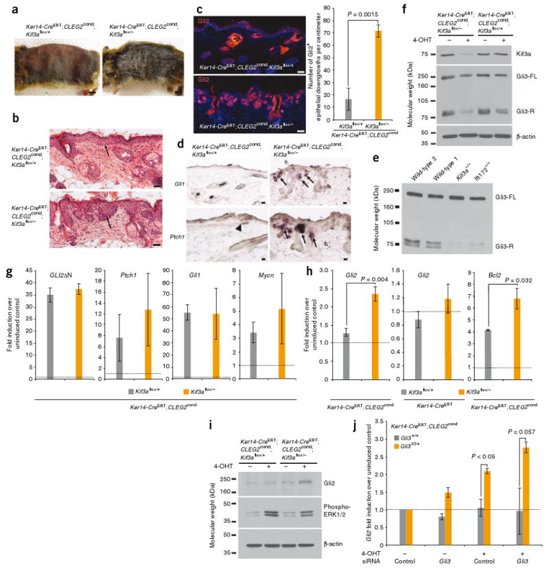
Primary cilia are present on most mammalian cells and are implicated in transducing Hedgehog (Hh) signals during development; however, the prevalence of cilia on human tumors remains unclear, and the role of cilia in cancer has not been examined. Here we show that human basal cell carcinomas (BCCs) are frequently ciliated, and we test the role of cilia in BCC by conditionally deleting Kif3a (encoding kinesin family member 3A) or Ift88 (encoding intraflagellar transport protein 88), genes required for ciliogenesis, in two Hh pathway-dependent mouse tumor models. Ciliary ablation strongly inhibited BCC-like tumors induced by an activated form of Smoothened. In contrast, removal of cilia accelerated tumors induced by activated Gli2, a transcriptional effector of Hh signaling. These seemingly paradoxical effects are consistent with a dual role for cilia in mediating both the activation and the repression of the Hh signaling pathway. Our findings demonstrate that cilia function as unique signaling organelles that can either mediate or suppress tumorigenesis depending on the nature of the oncogenic initiating event.


Figures

Figure 1
Figure 1
BCC and normal skin cells possess primary cilia. (a) Immunofluorescence imaging of the ciliary marker acetylated tubulin (green) and rootletin (red), a ciliary rootlet component. Five of eight independent human clinical BCCs, including nodular and superficial subtypes, contain numerous ciliated cells (arrows). Insets, H&E staining of adjacent biopsies. Right images are views of the left and middle panels. (b) Immunofluorescence imaging of the ciliary marker detyrosinated tubulin (green) and the centrosomal marker γ -tubulin (red) (dotted line, epidermal basal layer;inset, enlarged view). Arrows indicate ciliated cells present in SmoM2-induced regions of mouse epidermal hyperplasia. (c) Cilia extending from normal skin cells (arrows). Left images, low magnification;right images, high magnification of boxed areas. (d) Cilia extending from cultured keratinocytes (arrows). All scale bars are 50 μm except that in d (10 μm).
Figure 2
Figure 2
Cilia are essential for SmoM2-induced neoplasia. (a) Schematic showing that skin epithelial cells exposed to tamoxifen (TAM) both delete Kif3aflox and activate SmoM2cond (red cells), whereas unexposed cells retain Kif3aflox and do not activate SmoM2cond (gray cells). Cilia are lost in cells from Kif3aflox/− but not Kif3aflox/+ mice exposed to tamoxifen. (b) H&E staining of dorsal skin biopsies, showing that removal of Kif3a (Kif3aflox/−) blocks SmoM2-initiated tumorigenesis (arrows). (c) Immunofluorescence imaging showing that SmoM2 localizes to cilia (arrows) in neoplastic epidermal downgrowths. (d) Quantification of interfollicular epidermis thickness. Seven or eight independent Ker 14-CreERT;SmoM2cond;Kif3aflox/+ and Ker14-CreERT;SmoM2cond;Kif3aflox/− mice per genotype were analyzed. Four or five mice were analyzed for all other genotypes. (e) Protection against SmoM2-induced ear skin hyperplasia upon loss of Kif3a (arrow). (f) H&E staining of representative ear sections. (g) In situ staining showing that SmoM2-induced skin lesions upregulate Hh target genes Gli1 and Ptch1 (arrows). (h) Immunofluorescence imaging showing that Gli2 is upregulated in SmoM2-induced tumors that express Kif3a (stainings are representative of the mice analyzed in d and j). (i) Immunofluorescence imaging showing that Gli2 is upregulated upon expression of Myc-tagged SmoA1 in ciliated (wild type), but not unciliated (Kif3a−/− and Ift172−/−), transformed MEFs. (j) Cellular proliferation in skin, as assessed by Ki67 staining. Eight Ker14-CreERT;SmoM2cond;Kif3aflox/+ and Ker14-CreERT;SmoM2cond;Kif3aflox/ mice per genotype were analyzed, as well as four or five mice for the other genotypes. All scale bars are 50 μm except that in e (5 mm). All error bars shown, ± s.e.m.
Figure 3
Figure 3
Loss of cilia accelerates GLI2ΔN-induced neoplasia. (a) Gross morphology of mice upon induction of CLEG2cond. (b) H&E staining showing that loss of Kif3a increases the severity of GLI2ΔN-induced skin lesions (arrows). Images are representative of five or six mice analyzed per genotype. (c) Immunofluorescence imaging showing that loss of Kif3a increases the number of Gli2+ downgrowths in the skin. (d) In situ staining revealing that Ptch1 is upregulated in small regions of CLEG2-expressing Kif3aflox/+ skin (arrowhead), whereas Gli1 and Ptch1 are more frequently upregulated in CLEG2-induced lesions lacking Kif3a (arrows). (e) Formation of Gli3-R in ciliated (wild type) and non-ciliated (Kif3a−/−, Ift172−/−) transformed MEFs (Gli3-FL, full length Gli3). (f) Western blot indicating that Kif3a protein expression is lower and Gli3-R formation is impaired in 4-OHT–treated Ker14-CreERT;CLEG2cond;Kif3aflox/−keratinocytes, relative to control cells. (g) Expression of constitutively active human GLI2 (GLI2ΔN), Ptch1, Gli1 and Mycn in Kif3aflox/+ and Kif3aflox/−keratinocytes treated with 4-OHT. (All expression values expressed relative to those of vehicle-treated control cells, set to a baseline of ‘1’, dotted line.) (h) Quantiative RT-PCR assessment of GLI2ΔN-mediated induction of endogenous Gli2 and Bcl2 in ciliated and unciliated Kif3aflox/− keratinocytes. (i) Endogenous Gli2 and phosphorylated ERK1/2 protein levels in 4-OHT–treated Ker14-CreERT;CLEG2cond keratinocytes that are either Kif3aflox/+ or Kif3aflox/−. (j) Expression of endogenous Gli2 in GLI2ΔN-induced Gli3Xt/+ or Gli3+/+ keratinocytes, treated with siRNA against Gli3 or control. All expression values are normalized to those of vehicle-treated cells transfected with control siRNAs (baseline set to ‘1’, dotted line). All scale bars are 50 μm except that in a (5 mm). All error bars show means ± s.e.m.
Figure 4
Figure 4
Loss of Ift88 restrains SmoM2-mediated tumorigenesis and promotes GLI2ΔN-induced BCC-like lesions in tamoxifen-treated mice. (a) H&E staining of dorsal skin biopsies, revealing that loss of Ift88 (Ift88flox/−) blocks SmoM2-induced epidermal hyperplasia and tumorigenesis. (b) Quantification of epidermal thickness for a. Three to five mice per genotype were analyzed 10 weeks after induction by tamoxifen, and five mice per genotype were assessed 20 weeks after tamoxifen treatment. (c) H&E staining revealing that loss of Ift88 promotes skin lesions induced by activated Gli2. All scale bars, 50 μm. (d) Quantification of the number of Gli2+ downgrowths in the skin. Four mice were analyzed per genotype. (e) Schematic showing that in nonpathological conditions and in the absence of Hh, the cilium mediates the formation of Gli3-R, a repressor of downstream Hh target genes. In the presence of Hh ligand, Ptch1-mediated inhibition of Smo is suppressed, allowing Smo to act through the cilium to inhibit Gli3-R formation and induce Gli activators (Gli-Act), which turn on the Hh transcriptional program. In cilium-dependent oncogenesis (for example, SmoM2-induced tumors), the cilium is essential for Gli-Act formation and, thus, tumorigenesis. In cilium-independent oncogenesis (for example, tumors induced by constitutively active GLI2 (Gli-Act*)), the cilium restrains tumorigenesis through production of counter-balancing Gli3-R. Loss of the cilium inhibits Gli3-R formation, allowing unopposed Gli-Act* to aggressively drive tumorigenesis. All error bars show means ± s.e.m.

Comment in

  • Two sides to cilia in cancer.
    Toftgård R. Toftgård R. Nat Med. 2009 Sep;15(9):994-6. doi: 10.1038/nm0909-994. Nat Med. 2009. PMID: 19734870 No abstract available.

References

    1. Dahmane N, Lee J, Robins P, Heller P, Ruiz i Altaba A. Activation of the transcription factor Gli1 and the Sonic hedgehog signalling pathway in skin tumours. Nature. 1997;389:876–881. - PubMed
    1. Rubin LL, de Sauvage FJ. Targeting the Hedgehog pathway in cancer. Nat Rev Drug Discov. 2006;5:1026–1033. - PubMed
    1. Hooper JE, Scott MP. Communicating with Hedgehogs. Nat Rev Mol Cell Biol. 2005;6:306–317. - PubMed
    1. Xie J, et al. Activating Smoothened mutations in sporadic basal-cell carcinoma. Nature. 1998;391:90–92. - PubMed
    1. Reifenberger J, et al. Missense mutations in SMOH in sporadic basal cell carcinomas of the skin and primitive neuroectodermal tumors of the central nervous system. Cancer Res. 1998;58:1798–1803. - PubMed

Publication types

MeSH terms