. 2009 Sep;30(9):1297-306.
doi: 10.1038/aps.2009.124. Epub 2009 Aug 24.

n-Butylidenephthalide induced apoptosis in the A549 human lung adenocarcinoma cell line by coupled down-regulation of AP-2alpha and telomerase activity

Affiliations

n-Butylidenephthalide induced apoptosis in the A549 human lung adenocarcinoma cell line by coupled down-regulation of AP-2alpha and telomerase activity

Chyou-wei Wei et al. Acta Pharmacol Sin. 2009 Sep.

Expression of concern in

Abstract

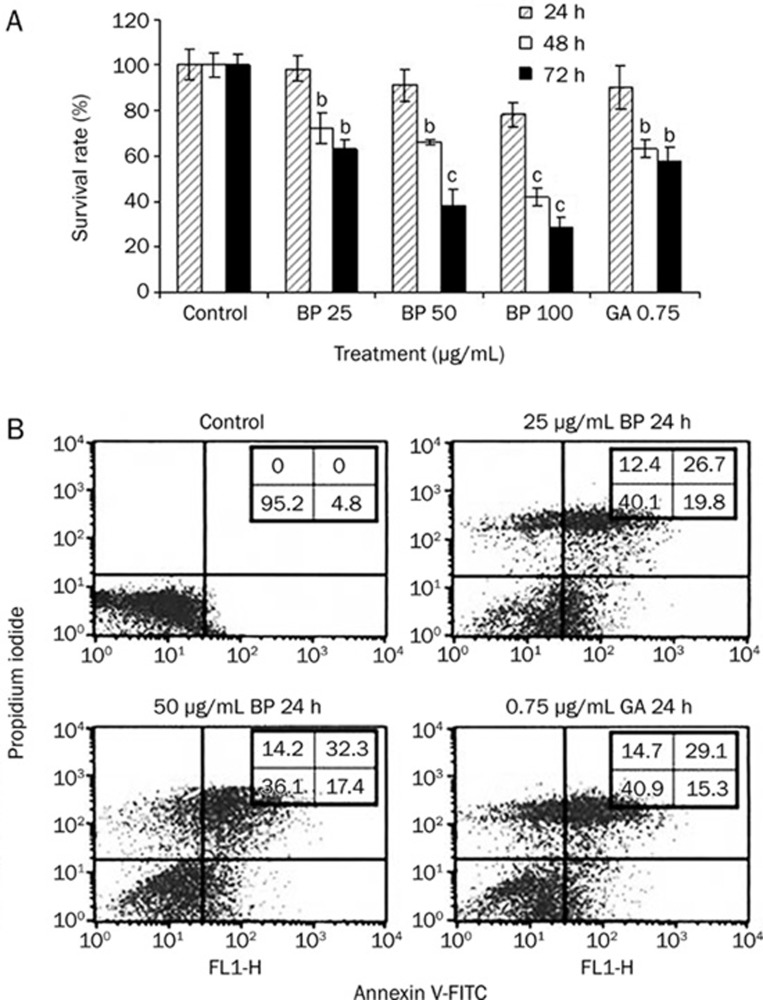
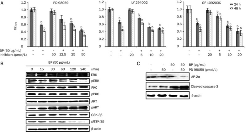
Aim: To investigate the role of hTERT gene expression and AP-2alpha in n-butylidenephthalide (n-BP)-induced apoptosis in A549 lung cancer cells.

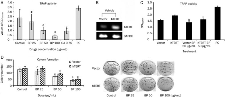
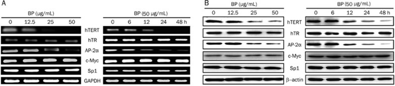
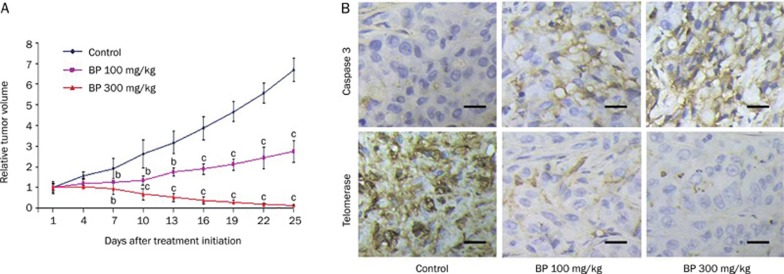
Methods: Viability of A549 cells was measured by MTT assay. Protein expression was determined by Western blot. Telomerase activity was measured using the modified telomere repeat amplification protocol (TRAP) assay. Xenograft mice were used as a model system to study the cytotoxic effect of n-BP in vivo. The morphology of tumor was examined by immunohistochemical staining.

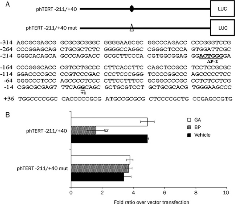
Results: The growth of A549 lung cancer cells treated with n-BP was significantly inhibited. Telomerase activity and hTERT mRNA expression were determined by telomeric repeat amplification protocol and reverse transcription-polymerase chain reaction, respectively. n-BP inhibited telomerase activity and hTERT mRNA expression in A549 cells while overexpression of hTERT could abolish BP-induced growth inhibition in the A549 cells. We also showed that hTERT promoter activity in the presence of n-BP was mediated via AP-2alpha. We saw an inhibition of tumor growth when nude mice carrying A549 subcutaneous xenograft tumors were treated with n-BP. Immunohistochemistry of this tumor tissue also showed a decrease in the expression of hTERT.

Conclusion: The antiproliferative effects of n-BP on A549 cells in vitro and in vivo suggest a novel clinical application of this compound in the treatment of lung cancers.


Figures

Figure 1
Figure 1
BP caused growth inhibition of human lung adenocarcinoma cells in vitro and induces apoptosis. (A) Growth inhibition effect was assessed in human lung cell lines A549. The cells were treated with various concentrations ranging from 25 to 100 μg/mL BP or vehicle (0.2% DMSO) for various times (24, 48, and 72 h) as indicated. GA-treated cells were used as positive control. Growth inhibition effect (IC50) was determined by MTT assay. (B) cells were treated with various concentrations of 0 to 50 μg/mL BP or vehicle for 24 h. Cells were collected, and apoptosis was analyzed using PI/Annexin V staining. The data represent the means±SD of three different experiments. bP<0.05, cP<0.01 vs the control.
Figure 2
Figure 2
Effects of BP treatment on telomerase activity in human lung cancer cell line A549. (A) Telomerase was extracted from A549 cells treated with BP at various concentrations for a period of 48 h. Telomerase activity was determined using the TRAP ELISA method as described in Materials and methods. Positive control (P.C.) represents assay with telomerase extract from kit, while DMSO is cells treated with solvent (0.1% DMSO). GA indicated Gambogic acid-treated A549 cells. BP dose-dependently inhibited telomerase activity in the A549 cell line. The data represent the means±SD of three different experiments. bP<0.05, cP<0.01 vs the control. (B) The full-length hTERT was cloned into pCI-neo5.4 expression vector and trnasfected into A549 cells. Each cell was grown under G418 (500 μg/mL) for 2 weeks. Total mRNA of 2 μg was loaded in each lane and RT-PCR analysis was described in “Materials and Methods”. (C) Telomerase activity in transfected A549 cells. A549 cells transfected with one of the pCI-neo5.4 (CI-neo5.4; empty vector) and pCI-neo-hEST2-HA8.9 (hTERT) and incubated in the presence of BP (50 μg/mL) for 48 h. Cells were collected and telomerase activity was analyzed using TRAP ELISA method. The data reveals that A549 cells contain more activity of hTERT than vector alone, and shown that BP could repress telomerase activity. The data represent the means±SD of three different experiments. bP<0.05, cP<0.01 vs the control. (D) Effects of hTERT expression on BP-induced apoptosis with colony formation. Colony formation assay using hTERT overexpression cells. A549 cells were transfected with either pCI-neo5.4 or pCI-neo-hEST2-HA8.9, and stably transfected clones were selected by exposoure to G418 (500 μg/mL) for 2 weeks. The stably transfected clones were treated with either vehicle or BP at various concentrations for a period of 48 h and then grown in culture medium for 2 weeks. The data represent the means±SD of three different experiments. bP<0.05, cP<0.01 vs the control.
Figure 3
Figure 3
Effect of n-BP on mRNA and protein expression of hTERT, hTR, AP-2α, c-Myc and Sp1 in A549 cells. (A) mRNA expression of hTERT, hTR, AP-2α, c-Myc and Sp1 in the cells treated with various concentrations of n-BP (0, 12.5, 25 and 50 μg/mL) for 2 days and the cells treated with 50 μg/mL BP or vehicle for various durations (0, 6, 12, 24, and 48 h) as indicated. Cells were collected and total RNA isolated for RT-PCR analysis, and expression of GAPDH was used as an internal control. (B) protein expressions of hTERT, hTR, AP-2α, c-Myc and Sp1 in the cells treated with various concentrations of n-BP (0, 12.5, 25 and 50 μg/mL) for 2 days and the cells treated with 50 μg/mL BP or vehicle for various durations (0, 6, 12, 24, and 48 h) as indicated. These expressions were assessed by Western blot assay, and expression of β-actin was used as an internal control.
Figure 4
Figure 4
The AP-2α binding site has an important role in BP-induced hTERT expression. (A) comparison of wild-type and mutant sequences between -211 and +40 in the hTERT promoter region. Mutated base pairs are indicated in each mutated sequence. (B) wildtype (phTERT -211/+40) or AP-2α mutant vector (phTERT -211/+40 mut) and pRL-null cotransfected into A549 cells. These indicated promoter regions were fused to a luciferase report gene. Each construct (2 μg) was cotransfected with 0.2 μg of pRL-null vector into A549 cells using GeneJammmer transfection reagent followed by treatment with 50 μg/mL BP for 48 h. Cells were lysed, and luciferase activity was measured. Transfection efficiency for luciferase activity was normalized to vehicle luciferase (pRL-null vector). The x-axis shows relative luciferase units (RLU) (firefly luciferase/vehicle luciferase). The data represent the means±SD of three different experiments. bP<0.05 vs the control.
Figure 5
Figure 5
Inhibition of ERK expression and enhanced growth inhibition by MEK inhibitor PD98059. A, MTT assay of A549 cells with culture or serum-containing medium pretreated with the MEK1/2 inhibitor PD98059 (12.5, 25, and 50 μmol/L), the PKC inhibitor GF109203X (5, 10, and 20 μmol/L), or the PI3K/AKT inhibitor LY294002 (5, 10, and 20 μmol/L) for 1 h and then treated with 50 μg/mL BP for 24 and 48 h. Lane 1 shows A549 cells treated with serum containing media and no test compound as a negative control. The data represent the means±SD of three different experiments. bP<0.05, cP<0.01 vs the vehicle. B, Western blot analysis of ERK, phosphor-ERK (pERK), PKC, phosphor-PKC (pPKC), AKT, phosphor-AKT (pAKT), GSK-3β, and phosphor-GSK-3β (pGSK-3β) in A549 cells after treatment with 50 μg/mL BP for the indicated times. The expression of β-actin was used as an internal control. (C) Inhibition of AP-2 and enhancement of cleaved caspase-3 expression by MEK inhibitor in the BP-induced growth inhibition. A549 cells were in incubated in the presence or absence of the MEK inhibitor PD98059 for 1 h and then treated with BP for 12 h. Western blot analysis was performed for AP-2α, cleaved caspase-3, and expression of β-actin was used as an internal control.
Figure 6
Figure 6
BP-induced in vivo growth inhibition of A549 xenografts in nude mice. Inhibition of tumor growth from A549 cells that downregulation of hTERT protein. Nude mice injected with approximately 5×10⁶ A549 into the dorsal subcutaneous tissue. (A) When the tumor volumes reached 80-120 mm3, A549 tumor-bearing mice were administered sc with vehicle control (♦), 100 mg/kg BP (▪), and 300 mg/kg n-BP (▴) on days 0–4 for 5 days. These figures shows A549 cells relative tumor volume of control and therapeutic groups. (B) immunohistochemical staining was analyzed in A549 tumor tissues (at day 10 after initiation of drug treatment). Representative photographs of sections of the control group and n-BP-treated group. A549 tumors, immunohistochemically stained with hTERT and caspase-3 rabbit polyclonal antibody, and hTERT and caspase-3 positive cells were stained brown (black arrowheads) (×400). Scale bars, 100 μm. n=3 different experiments. Means±SD. bP<0.05, cP<0.01 vs the control.

References

    1. Blackburn EH. Telomerases. Annu Rev Biochem. 1992;61:113–29. - PubMed
    1. Greider CW, Blackburn EH. A telomeric sequence in the RNA of tetrahymena telomerase required for telomere repeat synthesis. Nature. 1989;337:331–7. - PubMed
    1. Blackburn EH. Structure and function of telomeres. Nature. 1991;350:569–73. - PubMed
    1. Feng J, Funk WD, Wang SS, Weinrich SL, Avilion AA, Chiu CP, et al. The RNA component of human telomerase. Science. 1995;269:1236–41. - PubMed
    1. Nakamura TM, Morin GB, Chapman KB, Weinrich SL, Andrews WH, Lingner J, et al. Telomerase catalytic subunit homologs from fission yeast and human. Science. 1997;277:955–9. - PubMed

Publication types

MeSH terms